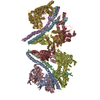
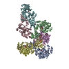
[日本語] English
- PDB-6lu9: Rat ionotropic Glutamate Delta-2 receptor in complex with 7-CKA a... -

+
データを開く


IDまたはキーワード:

読み込み中...

-
基本情報

登録情報
データベース: PDB / ID: 6lu9
タイトルRat ionotropic Glutamate Delta-2 receptor in complex with 7-CKA and Calcium
要素Glutamate receptor ionotropic, delta-2
キーワードMEMBRANE PROTEIN
機能・相同性
機能・相同性情報


excitatory synapse assembly / cerebellar granule cell differentiation / positive regulation of long-term synaptic depression / regulation of postsynaptic density assembly / glutamate receptor activity / positive regulation of synapse assembly / parallel fiber to Purkinje cell synapse / heterophilic cell-cell adhesion via plasma membrane cell adhesion molecules / regulation of neuron projection development / ionotropic glutamate receptor complex ...excitatory synapse assembly / cerebellar granule cell differentiation / positive regulation of long-term synaptic depression / regulation of postsynaptic density assembly / glutamate receptor activity / positive regulation of synapse assembly / parallel fiber to Purkinje cell synapse / heterophilic cell-cell adhesion via plasma membrane cell adhesion molecules / regulation of neuron projection development / ionotropic glutamate receptor complex / regulation of presynapse assembly / prepulse inhibition / regulation of neuron apoptotic process / somatodendritic compartment / excitatory postsynaptic potential / synaptic transmission, glutamatergic / transmitter-gated monoatomic ion channel activity involved in regulation of postsynaptic membrane potential / PDZ domain binding / postsynaptic density membrane / modulation of chemical synaptic transmission / protein localization / scaffold protein binding / postsynaptic membrane / dendritic spine / glutamatergic synapse / synapse / identical protein binding / membrane / plasma membrane
類似検索 - 分子機能
Ionotropic glutamate receptor, metazoa / Ligated ion channel L-glutamate- and glycine-binding site / : / Ligand-gated ion channel / Ionotropic glutamate receptor, L-glutamate and glycine-binding domain / Ligated ion channel L-glutamate- and glycine-binding site / Ionotropic glutamate receptor / Eukaryotic homologues of bacterial periplasmic substrate binding proteins. / Receptor, ligand binding region / Receptor family ligand binding region / Periplasmic binding protein-like I
類似検索 - ドメイン・相同性
Glutamate receptor ionotropic, delta-2
類似検索 - 構成要素
生物種Rattus norvegicus (ドブネズミ)
手法電子顕微鏡法 / 単粒子再構成法 / クライオ電子顕微鏡法 / 解像度: 8.8 Å
データ登録者Kumar, J. / Burada, A.P.
資金援助 インド, 1件
組織認可番号
Wellcome TrustIA/I/13/2/5010023 インド
引用ジャーナル: J Struct Biol / : 2020
タイトル: The architecture of GluD2 ionotropic delta glutamate receptor elucidated by cryo-EM.
著者: Ananth Prasad Burada / Rajesh Vinnakota / Janesh Kumar /
要旨: GluD2 receptor belongs to the orphan delta family of glutamate receptor ion channels. These receptors play key roles in synaptogenesis and synaptic plasticity and are associated with multiple ...GluD2 receptor belongs to the orphan delta family of glutamate receptor ion channels. These receptors play key roles in synaptogenesis and synaptic plasticity and are associated with multiple neuronal disorders like schizophrenia, autism spectrum disorder, cerebellar ataxia, intellectual disability, paraplegia, retinal dystrophy, etc. Despite the importance of these receptors in CNS, insights into full-length GluD2 receptor structure is missing till-date. Here we report cryo-electron microscopy structure of the rat GluD2 receptor in the presence of calcium ions and the ligand 7-chlorokynurenic acid, elucidating its 3D architecture. The structure reveals a non-swapped architecture at the extracellular amino-terminal (ATD), and ligand-binding domain (LBD) interface similar to that observed in GluD1; however, the organization and arrangement of the ATD and LBD domains in GluD2 are unique. While our results demonstrate that non-swapped architecture is conserved in the delta receptor family, they also highlight the differences that exist between the two member receptors; GluD1 and GluD2.
履歴
登録2020年1月27日登録サイト: PDBJ / 処理サイト: PDBJ
改定 1.02020年6月17日Provider: repository / タイプ: Initial release
改定 1.12020年12月30日Group: Database references / カテゴリ: citation / citation_author
Item: _citation.country / _citation.journal_abbrev ..._citation.country / _citation.journal_abbrev / _citation.journal_id_ASTM / _citation.journal_id_CSD / _citation.journal_id_ISSN / _citation.journal_volume / _citation.page_first / _citation.page_last / _citation.pdbx_database_id_DOI / _citation.pdbx_database_id_PubMed / _citation.title / _citation.year

-
構造の表示

ムービー
  • 登録構造単位
  • Jmolによる作画
  • ダウンロード
  • 単純化した表面モデル + あてはめた原子モデル
  • マップデータ: EMDB-0979
  • Jmolによる作画
  • ダウンロード
  • EMマップとの重ね合わせ
  • マップデータ: EMDB-0979
  • UCSF Chimeraによる作画
  • ダウンロード
ムービービューア
構造ビューア分子:
MolmilJmol/JSmol

ダウンロードとリンク

-
集合体

登録構造単位
A: Glutamate receptor ionotropic, delta-2
B: Glutamate receptor ionotropic, delta-2
C: Glutamate receptor ionotropic, delta-2
D: Glutamate receptor ionotropic, delta-2


分子量 (理論値)分子数
合計 (水以外)394,7694
ポリマ-394,7694
非ポリマー00
00
1


  • 登録構造と同一
  • 登録者が定義した集合体
  • 根拠: gel filtration
タイプ名称対称操作
identity operation1_5551
Buried area8360 Å2
ΔGint-22 kcal/mol
Surface area116920 Å2

-
要素

#1: タンパク質
Glutamate receptor ionotropic, delta-2 / GluR delta-2 subunit


分子量: 98692.242 Da / 分子数: 4 / 由来タイプ: 組換発現 / 由来: (組換発現) Rattus norvegicus (ドブネズミ) / 遺伝子: Grid2 / プラスミド: pEGBacMam / 発現宿主: 
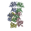
Homo sapiens (ヒト) / 参照: UniProt: Q63226

-
実験情報

-
実験

実験手法: 電子顕微鏡法
EM実験試料の集合状態: PARTICLE / 3次元再構成法: 単粒子再構成法

-
試料調製

構成要素名称: GluD2 / タイプ: COMPLEX / 詳細: Recombinantly expressed protein / Entity ID: all / 由来: RECOMBINANT
分子量: 0.388 MDa / 実験値: YES
由来(天然)生物種: Rattus norvegicus (ドブネズミ)
由来(組換発現)生物種: Homo sapiens (ヒト) / 細胞: HEK293S GnTI- / プラスミド: pEG Bac Mam
緩衝液pH: 8 / 詳細: 20mM Tris, 150mM NaCl, 0.75mM DDM, 0.025mM CHS
試料濃度: 0.9 mg/ml / 包埋: NO / シャドウイング: NO / 染色: NO / 凍結: YES
急速凍結装置: FEI VITROBOT MARK IV / 凍結剤: ETHANE / 湿度: 95 % / 凍結前の試料温度: 277 K

-
電子顕微鏡撮影

実験機器
モデル: Titan Krios / 画像提供: FEI Company
顕微鏡モデル: FEI TITAN KRIOS
電子銃電子線源: FIELD EMISSION GUN / 加速電圧: 300 kV / 照射モード: FLOOD BEAM
電子レンズモード: BRIGHT FIELD / Cs: 2.7 mm
試料ホルダ凍結剤: NITROGEN
撮影平均露光時間: 6 sec. / 電子線照射量: 40.38 e/Å2
フィルム・検出器のモデル: GATAN K2 SUMMIT (4k x 4k)
実像数: 4120
画像スキャン動画フレーム数/画像: 40 / 利用したフレーム数/画像: 0-40

-
解析

ソフトウェア名称: PHENIX / バージョン: 1.17.1_3660: / 分類: 精密化
EMソフトウェア
ID名称バージョンカテゴリ詳細
2EPU画像取得FEI EPU
4Gctfv1.06CTF補正Gctf
7UCSF Chimera1.12モデルフィッティング
9PHENIX1.16モデル精密化
10cryoSPARCv2.13初期オイラー角割当
11cryoSPARCv2.13最終オイラー角割当local refinement
12cryoSPARCv2.13分類
13cryoSPARCv2.133次元再構成
CTF補正タイプ: PHASE FLIPPING AND AMPLITUDE CORRECTION
粒子像の選択選択した粒子像数: 61332 / 詳細: Particles were picked manually
対称性点対称性: C1 (非対称)
3次元再構成解像度: 8.8 Å / 解像度の算出法: FSC 0.143 CUT-OFF / 粒子像の数: 7977 / クラス平均像の数: 1 / 対称性のタイプ: POINT
原子モデル構築プロトコル: RIGID BODY FIT / 空間: REAL
拘束条件
Refine-IDタイプDev ideal
ELECTRON MICROSCOPYf_bond_d0.02421416
ELECTRON MICROSCOPYf_angle_d2.53929020
ELECTRON MICROSCOPYf_dihedral_angle_d22.4447920
ELECTRON MICROSCOPYf_chiral_restr0.1443216
ELECTRON MICROSCOPYf_plane_restr0.0193792

+
万見について

-
お知らせ

-
2022年2月9日: EMDBエントリの付随情報ファイルのフォーマットが新しくなりました

EMDBエントリの付随情報ファイルのフォーマットが新しくなりました

  • EMDBのヘッダファイルのバージョン3が、公式のフォーマットとなりました。
  • これまでは公式だったバージョン1.9は、アーカイブから削除されます。

関連情報:EMDBヘッダ

外部リンク:wwPDBはEMDBデータモデルのバージョン3へ移行します

-
2020年8月12日: 新型コロナ情報

新型コロナ情報

URL: https://pdbjlvh1.pdbj.org/emnavi/covid19.php

新ページ: EM Navigatorに新型コロナウイルスの特設ページを開設しました。

関連情報:Covid-19情報 / 2020年3月5日: 新型コロナウイルスの構造データ

+
2020年3月5日: 新型コロナウイルスの構造データ

新型コロナウイルスの構造データ

関連情報:万見生物種 / 2020年8月12日: 新型コロナ情報

外部リンク:COVID-19特集ページ - PDBj / 今月の分子2020年2月:コロナウイルスプロテーアーゼ

+
2019年1月31日: EMDBのIDの桁数の変更

EMDBのIDの桁数の変更

  • EMDBエントリに付与されているアクセスコード(EMDB-ID)は4桁の数字(例、EMD-1234)でしたが、間もなく枯渇します。これまでの4桁のID番号は4桁のまま変更されませんが、4桁の数字を使い切った後に発行されるIDは5桁以上の数字(例、EMD-12345)になります。5桁のIDは2019年の春頃から発行される見通しです。
  • EM Navigator/万見では、接頭語「EMD-」は省略されています。

関連情報:Q: 「EMD」とは何ですか? / 万見/EM NavigatorにおけるID/アクセスコードの表記

外部リンク:EMDB Accession Codes are Changing Soon! / PDBjへお問い合わせ

+
2017年7月12日: PDB大規模アップデート

PDB大規模アップデート

  • 新バージョンのPDBx/mmCIF辞書形式に基づくデータがリリースされました。
  • 今回の更新はバージョン番号が4から5になる大規模なもので、全エントリデータの書き換えが行われる「Remediation」というアップデートに該当します。
  • このバージョンアップで、電子顕微鏡の実験手法に関する多くの項目の書式が改定されました(例:em_softwareなど)。
  • EM NavigatorとYorodumiでも、この改定に基づいた表示内容になります。

外部リンク:wwPDB Remediation / OneDepデータ基準に準拠した、より強化された内容のモデル構造ファイルが、PDBアーカイブで公開されました。

-
万見 (Yorodumi)

幾万の構造データを、幾万の視点から

  • 万見(Yorodumi)は、EMDB/PDB/SASBDBなどの構造データを閲覧するためのページです。
  • EM Navigatorの詳細ページの後継、Omokage検索のフロントエンドも兼ねています。

関連情報:EMDB / PDB / SASBDB / 3つのデータバンクの比較 / 万見検索 / 2016年8月31日: 新しいEM Navigatorと万見 / 万見文献 / Jmol/JSmol / 機能・相同性情報 / 新しいEM Navigatorと万見の変更点

他の情報も見る