 Movie
Movie Controller

[English] 日本語
 Yorodumi
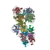
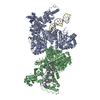
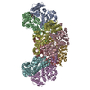
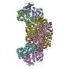
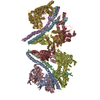
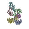
- PDB-6lu9: Rat ionotropic Glutamate Delta-2 receptor in complex with 7-CKA a... -
+ Open data

- Basic information
| Entry | Database: PDB / ID: 6lu9 | ||||||
|---|---|---|---|---|---|---|---|
| Title | Rat ionotropic Glutamate Delta-2 receptor in complex with 7-CKA and Calcium | ||||||
 Components Components | Glutamate receptor ionotropic, delta-2 | ||||||
 Keywords Keywords | MEMBRANE PROTEIN | ||||||
| Function / homology |  Function and homology information Function and homology informationtrans-synaptic protein complex / cerebellar granule cell differentiation / positive regulation of long-term synaptic depression / excitatory synapse assembly / synaptic signaling via neuropeptide / regulation of postsynaptic density assembly / positive regulation of synapse assembly / heterophilic cell-cell adhesion / regulation of neuron projection development / AMPA glutamate receptor activity ...trans-synaptic protein complex / cerebellar granule cell differentiation / positive regulation of long-term synaptic depression / excitatory synapse assembly / synaptic signaling via neuropeptide / regulation of postsynaptic density assembly / positive regulation of synapse assembly / heterophilic cell-cell adhesion / regulation of neuron projection development / AMPA glutamate receptor activity / parallel fiber to Purkinje cell synapse / AMPA glutamate receptor complex / ionotropic glutamate receptor complex / regulation of presynapse assembly / regulation of postsynaptic membrane neurotransmitter receptor levels / prepulse inhibition / somatodendritic compartment / regulation of neuron apoptotic process / PDZ domain binding / synaptic transmission, glutamatergic / transmitter-gated monoatomic ion channel activity involved in regulation of postsynaptic membrane potential / excitatory postsynaptic potential / postsynaptic density membrane / modulation of chemical synaptic transmission / intracellular protein localization / scaffold protein binding / dendritic spine / postsynaptic membrane / synapse / glutamatergic synapse / membrane / identical protein binding / plasma membrane Similarity search - Function | ||||||
| Biological species |  | ||||||
| Method | ELECTRON MICROSCOPY / single particle reconstruction / cryo EM / Resolution: 8.8 Å | ||||||
 Authors Authors | Kumar, J. / Burada, A.P. | ||||||
| Funding support |  India, 1items India, 1items
| ||||||
 Citation Citation | Journal: J Struct Biol / Year: 2020 Title: The architecture of GluD2 ionotropic delta glutamate receptor elucidated by cryo-EM. Authors: Ananth Prasad Burada / Rajesh Vinnakota / Janesh Kumar / Abstract: GluD2 receptor belongs to the orphan delta family of glutamate receptor ion channels. These receptors play key roles in synaptogenesis and synaptic plasticity and are associated with multiple ...GluD2 receptor belongs to the orphan delta family of glutamate receptor ion channels. These receptors play key roles in synaptogenesis and synaptic plasticity and are associated with multiple neuronal disorders like schizophrenia, autism spectrum disorder, cerebellar ataxia, intellectual disability, paraplegia, retinal dystrophy, etc. Despite the importance of these receptors in CNS, insights into full-length GluD2 receptor structure is missing till-date. Here we report cryo-electron microscopy structure of the rat GluD2 receptor in the presence of calcium ions and the ligand 7-chlorokynurenic acid, elucidating its 3D architecture. The structure reveals a non-swapped architecture at the extracellular amino-terminal (ATD), and ligand-binding domain (LBD) interface similar to that observed in GluD1; however, the organization and arrangement of the ATD and LBD domains in GluD2 are unique. While our results demonstrate that non-swapped architecture is conserved in the delta receptor family, they also highlight the differences that exist between the two member receptors; GluD1 and GluD2. | ||||||
| History |
|
- Structure visualization
| Movie |
Movie viewer |
|---|---|
| Structure viewer | Molecule: MolmilJmol/JSmol |
- Downloads & links
-Download
| PDBx/mmCIF format | 6lu9.cif.gz | 471.1 KB | Display | PDBx/mmCIF format |
|---|---|---|---|---|
| PDB format | pdb6lu9.ent.gz | 368.7 KB | Display | PDB format |
| PDBx/mmJSON format | 6lu9.json.gz | Tree view | PDBx/mmJSON format | |
| Others |  Other downloads Other downloads |
-Validation report
| Arichive directory | https://data.pdbj.org/pub/pdb/validation_reports/lu/6lu9ftp://data.pdbj.org/pub/pdb/validation_reports/lu/6lu9 | HTTPS FTP |
|---|
-Related structure data
| Related structure data |  0979MC M: map data used to model this data C: citing same article ( |
|---|---|
| Similar structure data |


-Links
 PDBj
PDBj

- Assembly
| Deposited unit | 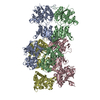
|
|---|---|
| 1 |
|
-Components
| #1: Protein | Mass: 98692.242 Da / Num. of mol.: 4 Source method: isolated from a genetically manipulated source Source: (gene. exp.)  Homo sapiens (human) / References: UniProt: Q63226 Homo sapiens (human) / References: UniProt: Q63226Has protein modification | Y | |
|---|
-Experimental details
-Experiment
| Experiment | Method: ELECTRON MICROSCOPY |
|---|---|
| EM experiment | Aggregation state: PARTICLE / 3D reconstruction method: single particle reconstruction |
- Sample preparation
| Component | Name: GluD2 / Type: COMPLEX / Details: Recombinantly expressed protein / Entity ID: all / Source: RECOMBINANT |
|---|---|
| Molecular weight | Value: 0.388 MDa / Experimental value: YES |
| Source (natural) | Organism: |
| Source (recombinant) | Organism: Homo sapiens (human) / Cell: HEK293S GnTI- / Plasmid: pEG Bac Mam |
| Buffer solution | pH: 8 / Details: 20mM Tris, 150mM NaCl, 0.75mM DDM, 0.025mM CHS |
| Specimen | Conc.: 0.9 mg/ml / Embedding applied: NO / Shadowing applied: NO / Staining applied: NO / Vitrification applied: YES |
| Vitrification | Instrument: FEI VITROBOT MARK IV / Cryogen name: ETHANE / Humidity: 95 % / Chamber temperature: 277 K |
- Electron microscopy imaging
| Experimental equipment |  Model: Titan Krios / Image courtesy: FEI Company |
|---|---|
| Microscopy | Model: FEI TITAN KRIOS |
| Electron gun | Electron source:  FIELD EMISSION GUN / Accelerating voltage: 300 kV / Illumination mode: FLOOD BEAM FIELD EMISSION GUN / Accelerating voltage: 300 kV / Illumination mode: FLOOD BEAM |
| Electron lens | Mode: BRIGHT FIELD / Cs: 2.7 mm |
| Specimen holder | Cryogen: NITROGEN |
| Image recording | Average exposure time: 6 sec. / Electron dose: 40.38 e/Å2 / Film or detector model: GATAN K2 SUMMIT (4k x 4k) / Num. of real images: 4120 |
| Image scans | Movie frames/image: 40 / Used frames/image: 0-40 |
- Processing
| Software | Name: PHENIX / Version: 1.17.1_3660: / Classification: refinement | |||||||||||||||||||||||||||||||||||||||||||||
|---|---|---|---|---|---|---|---|---|---|---|---|---|---|---|---|---|---|---|---|---|---|---|---|---|---|---|---|---|---|---|---|---|---|---|---|---|---|---|---|---|---|---|---|---|---|---|
| EM software |
| |||||||||||||||||||||||||||||||||||||||||||||
| CTF correction | Type: PHASE FLIPPING AND AMPLITUDE CORRECTION | |||||||||||||||||||||||||||||||||||||||||||||
| Particle selection | Num. of particles selected: 61332 / Details: Particles were picked manually | |||||||||||||||||||||||||||||||||||||||||||||
| Symmetry | Point symmetry: C1 (asymmetric) | |||||||||||||||||||||||||||||||||||||||||||||
| 3D reconstruction | Resolution: 8.8 Å / Resolution method: FSC 0.143 CUT-OFF / Num. of particles: 7977 / Num. of class averages: 1 / Symmetry type: POINT | |||||||||||||||||||||||||||||||||||||||||||||
| Atomic model building | Protocol: RIGID BODY FIT / Space: REAL | |||||||||||||||||||||||||||||||||||||||||||||
| Refine LS restraints |
|
