 Movie
Movie Controller


+ Open data

- Basic information
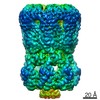
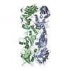
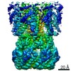
| Entry | Database: PDB / ID: 6kz3 | |||||||||
|---|---|---|---|---|---|---|---|---|---|---|
| Title | YebT domain1-4 | |||||||||
 Components Components | Intermembrane transport protein YebT | |||||||||
 Keywords Keywords | LIPID TRANSPORT / lipid channel | |||||||||
| Function / homology | : / Mce/MlaD / MlaD protein / intermembrane lipid transfer / membrane organization / outer membrane-bounded periplasmic space / identical protein binding / plasma membrane / Lipophilic envelope-spanning tunnel protein B Function and homology information Function and homology information | |||||||||
| Biological species |  | |||||||||
| Method | ELECTRON MICROSCOPY / single particle reconstruction / cryo EM / Resolution: 3.1 Å | |||||||||
 Authors Authors | Wang, H.W. / Liu, C. / Zhang, L. | |||||||||
| Funding support |  China, 2items China, 2items
| |||||||||
 Citation Citation | Journal: J Mol Biol / Year: 2020 Title: Cryo-EM Structure of a Bacterial Lipid Transporter YebT. Authors: Chuan Liu / Jinying Ma / Jia Wang / Hongwei Wang / Li Zhang / Abstract: The outer membrane (OM) of Gram-negative bacteria is asymmetric, with lipopolysaccharides (LPSs) on the outer surface and phospholipids (PLs) on the inner surface. This unique organization of OM ...The outer membrane (OM) of Gram-negative bacteria is asymmetric, with lipopolysaccharides (LPSs) on the outer surface and phospholipids (PLs) on the inner surface. This unique organization of OM makes Gram-negative bacteria resistant to many toxic chemicals. How this asymmetric distribution of lipids is maintained has been studied for decades with previous reports of an Mla (Maintenance of OM Lipid Asymmetry) system to be involved. Furthermore, the OM of Gram-negative bacteria is about 20 nm away from inner membrane (IM) where the lipids are synthesized. Therefore, how nascent lipids travel across the periplasmic space and arrive at the inner surface of OM is another interesting question. YebT is a homologue of MlaD in the Mla pathway, but its role in lipid distribution of the OM and IM is largely unknown. Here we report the first high-resolution (~3.0 Å) cryo-EM structure of full-length E. coli YebT in a substrate-bound state. Our structure with details of lipid interaction indicates that YebT is a lipid transporter spanning between IM and OM. We also demonstrate the symmetry mismatch in YebT and the existence of many other conformations of YebT revealing the intrinsic dynamics of this lipid channel. And a brief discussion on possible mechanisms of lipid transport is also included. | |||||||||
| History |
|
- Structure visualization
| Movie |
Movie viewer |
|---|---|
| Structure viewer | Molecule: MolmilJmol/JSmol |
- Downloads & links
-Download
| PDBx/mmCIF format | 6kz3.cif.gz | 433.7 KB | Display | PDBx/mmCIF format |
|---|---|---|---|---|
| PDB format | pdb6kz3.ent.gz | 359.8 KB | Display | PDB format |
| PDBx/mmJSON format | 6kz3.json.gz | Tree view | PDBx/mmJSON format | |
| Others |  Other downloads Other downloads |
-Validation report
| Arichive directory | https://data.pdbj.org/pub/pdb/validation_reports/kz/6kz3ftp://data.pdbj.org/pub/pdb/validation_reports/kz/6kz3 | HTTPS FTP |
|---|
-Related structure data
| Related structure data |  0784MC  0785C 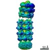 0786C  0787C  0788C  6kz4C M: map data used to model this data C: citing same article ( |
|---|---|
| Similar structure data |

-Links
 PDBj
PDBj- Assembly
| Deposited unit | 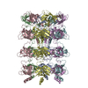
|
|---|---|
| 1 |
|
-Components
| #1: Protein | Mass: 50016.824 Da / Num. of mol.: 6 / Fragment: domain 1-4 Source method: isolated from a genetically manipulated source Source: (gene. exp.) |
|---|
-Experimental details
-Experiment
| Experiment | Method: ELECTRON MICROSCOPY |
|---|---|
| EM experiment | Aggregation state: PARTICLE / 3D reconstruction method: single particle reconstruction |
- Sample preparation
| Component | Name: YebT-N / Type: COMPLEX / Details: Domain 1-4 of YebT / Entity ID: all / Source: RECOMBINANT |
|---|---|
| Molecular weight | Value: 0.3 MDa / Experimental value: NO |
| Source (natural) | Organism: |
| Source (recombinant) | Organism: |
| Buffer solution | pH: 8 |
| Specimen | Embedding applied: NO / Shadowing applied: NO / Staining applied: NO / Vitrification applied: YES |
| Vitrification | Cryogen name: ETHANE |
- Electron microscopy imaging
| Experimental equipment |  Model: Titan Krios / Image courtesy: FEI Company |
|---|---|
| Microscopy | Model: FEI TITAN KRIOS |
| Electron gun | Electron source:  FIELD EMISSION GUN / Accelerating voltage: 300 kV / Illumination mode: FLOOD BEAM FIELD EMISSION GUN / Accelerating voltage: 300 kV / Illumination mode: FLOOD BEAM |
| Electron lens | Mode: BRIGHT FIELD |
| Image recording | Electron dose: 50 e/Å2 / Detector mode: SUPER-RESOLUTION / Film or detector model: GATAN K2 SUMMIT (4k x 4k) |
- Processing
| Software | Name: PHENIX / Version: 1.16_3549: / Classification: refinement | ||||||||||||||||||||||||
|---|---|---|---|---|---|---|---|---|---|---|---|---|---|---|---|---|---|---|---|---|---|---|---|---|---|
| CTF correction | Type: PHASE FLIPPING AND AMPLITUDE CORRECTION | ||||||||||||||||||||||||
| Symmetry | Point symmetry: C6 (6 fold cyclic) | ||||||||||||||||||||||||
| 3D reconstruction | Resolution: 3.1 Å / Resolution method: FSC 0.143 CUT-OFF / Num. of particles: 194342 / Symmetry type: POINT | ||||||||||||||||||||||||
| Refine LS restraints |
|
