 Movie
Movie Controller


+ Open data

- Basic information
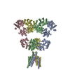
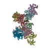
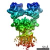
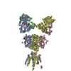
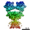
| Entry | Database: PDB / ID: 6jfz | |||||||||
|---|---|---|---|---|---|---|---|---|---|---|
| Title | GluK3 receptor complex with UBP310 | |||||||||
 Components Components | Glutamate receptor ionotropic, kainate 3 | |||||||||
 Keywords Keywords | MEMBRANE PROTEIN / Glutamate receptor / Kainate / UBP310 | |||||||||
| Function / homology |  Function and homology information Function and homology informationPresynaptic function of Kainate receptors / cochlear hair cell ribbon synapse / adenylate cyclase inhibiting G protein-coupled glutamate receptor activity / G protein-coupled glutamate receptor signaling pathway / Activation of Ca-permeable Kainate Receptor / kainate selective glutamate receptor complex / negative regulation of synaptic transmission, glutamatergic / glutamate receptor activity / glutamate receptor signaling pathway / kainate selective glutamate receptor activity ...Presynaptic function of Kainate receptors / cochlear hair cell ribbon synapse / adenylate cyclase inhibiting G protein-coupled glutamate receptor activity / G protein-coupled glutamate receptor signaling pathway / Activation of Ca-permeable Kainate Receptor / kainate selective glutamate receptor complex / negative regulation of synaptic transmission, glutamatergic / glutamate receptor activity / glutamate receptor signaling pathway / kainate selective glutamate receptor activity / glutamate-gated receptor activity / glutamate-gated calcium ion channel activity / dendrite cytoplasm / ligand-gated monoatomic ion channel activity involved in regulation of presynaptic membrane potential / synaptic transmission, glutamatergic / transmitter-gated monoatomic ion channel activity involved in regulation of postsynaptic membrane potential / regulation of membrane potential / postsynaptic density membrane / modulation of chemical synaptic transmission / terminal bouton / presynaptic membrane / chemical synaptic transmission / perikaryon / postsynaptic membrane / axon / dendrite / glutamatergic synapse / plasma membrane Similarity search - Function | |||||||||
| Biological species |  | |||||||||
| Method | ELECTRON MICROSCOPY / single particle reconstruction / cryo EM / Resolution: 7.6 Å | |||||||||
 Authors Authors | Kumari, J. / Kumar, J. | |||||||||
| Funding support |  India, 2items India, 2items
| |||||||||
 Citation Citation | Journal: Sci Rep / Year: 2019 Title: Structural and Functional Insights into GluK3-kainate Receptor Desensitization and Recovery. Authors: Jyoti Kumari / Rajesh Vinnakota / Janesh Kumar / Abstract: GluK3-kainate receptors are atypical members of the iGluR family that reside at both the pre- and postsynapse and play a vital role in the regulation of synaptic transmission. For a better ...GluK3-kainate receptors are atypical members of the iGluR family that reside at both the pre- and postsynapse and play a vital role in the regulation of synaptic transmission. For a better understanding of structural changes that underlie receptor functions, GluK3 receptors were trapped in desensitized and resting/closed states and structures analyzed using single particle cryo-electron microscopy. While the desensitized GluK3 has domain organization as seen earlier for another kainate receptor-GluK2, antagonist bound GluK3 trapped a resting state with only two LBD domains in dimeric arrangement necessary for receptor activation. Using structures as a guide, we show that the N-linked glycans at the interface of GluK3 ATD and LBD likely mediate inter-domain interactions and attune receptor-gating properties. The mutational analysis also identified putative N-glycan interacting residues. Our results provide a molecular framework for understanding gating properties unique to GluK3 and exploring the role of N-linked glycosylation in their modulation. | |||||||||
| History |
|
- Structure visualization
| Movie |
Movie viewer |
|---|---|
| Structure viewer | Molecule: MolmilJmol/JSmol |
- Downloads & links
-Download
| PDBx/mmCIF format | 6jfz.cif.gz | 498.3 KB | Display | PDBx/mmCIF format |
|---|---|---|---|---|
| PDB format | pdb6jfz.ent.gz | 415.4 KB | Display | PDB format |
| PDBx/mmJSON format | 6jfz.json.gz | Tree view | PDBx/mmJSON format | |
| Others |  Other downloads Other downloads |
-Validation report
| Arichive directory | https://data.pdbj.org/pub/pdb/validation_reports/jf/6jfzftp://data.pdbj.org/pub/pdb/validation_reports/jf/6jfz | HTTPS FTP |
|---|
-Related structure data
| Related structure data |  9822MC 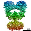 9821C  6jfyC  6jmvC C: citing same article ( M: map data used to model this data |
|---|---|
| Similar structure data |

-Links
 PDBj
PDBj

- Assembly
| Deposited unit | 
|
|---|---|
| 1 |
|
-Components
| #1: Protein | Mass: 91305.812 Da / Num. of mol.: 4 / Mutation: C86T, C305T, C547V Source method: isolated from a genetically manipulated source Source: (gene. exp.)  Homo sapiens (human) / References: UniProt: P42264 Homo sapiens (human) / References: UniProt: P42264Has protein modification | Y | Sequence details | The sequence conflicts A259P/S310P/P324A are based on AAC80577 (PubMed:1371217) according to ...The sequence conflicts A259P/S310P/P324A are based on AAC80577 (PubMed:1371217) according to database P42264 (GRIK3_RAT). | |
|---|
-Experimental details
-Experiment
| Experiment | Method: ELECTRON MICROSCOPY |
|---|---|
| EM experiment | Aggregation state: PARTICLE / 3D reconstruction method: single particle reconstruction |
- Sample preparation
| Component | Name: GluK3 complex with agonist SYM / Type: COMPLEX / Entity ID: all / Source: RECOMBINANT | |||||||||||||||||||||||||
|---|---|---|---|---|---|---|---|---|---|---|---|---|---|---|---|---|---|---|---|---|---|---|---|---|---|---|
| Molecular weight | Value: 0.4 MDa / Experimental value: NO | |||||||||||||||||||||||||
| Source (natural) | Organism: | |||||||||||||||||||||||||
| Source (recombinant) | Organism: Homo sapiens (human) / Cell: HEK293 / Plasmid: pEG BacMAM | |||||||||||||||||||||||||
| Buffer solution | pH: 8 | |||||||||||||||||||||||||
| Buffer component |
| |||||||||||||||||||||||||
| Specimen | Conc.: 1.7 mg/ml / Embedding applied: NO / Shadowing applied: NO / Staining applied: NO / Vitrification applied: YES / Details: Purified and detergent solubilized GluK3 receptors | |||||||||||||||||||||||||
| Vitrification | Cryogen name: ETHANE |
- Electron microscopy imaging
| Experimental equipment |  Model: Titan Krios / Image courtesy: FEI Company |
|---|---|
| Microscopy | Model: FEI TITAN KRIOS |
| Electron gun | Electron source:  FIELD EMISSION GUN / Accelerating voltage: 300 kV / Illumination mode: SPOT SCAN FIELD EMISSION GUN / Accelerating voltage: 300 kV / Illumination mode: SPOT SCAN |
| Electron lens | Mode: BRIGHT FIELD / Cs: 2 mm |
| Specimen holder | Cryogen: NITROGEN / Specimen holder model: FEI TITAN KRIOS AUTOGRID HOLDER |
| Image recording | Average exposure time: 60 sec. / Electron dose: 16.73 e/Å2 / Detector mode: COUNTING / Film or detector model: FEI FALCON III (4k x 4k) / Num. of grids imaged: 1 / Num. of real images: 719 |
- Processing
| Software | Name: PHENIX / Version: 1.14_3260: / Classification: refinement | ||||||||||||||||||||||||||||||||||||||||
|---|---|---|---|---|---|---|---|---|---|---|---|---|---|---|---|---|---|---|---|---|---|---|---|---|---|---|---|---|---|---|---|---|---|---|---|---|---|---|---|---|---|
| EM software |
| ||||||||||||||||||||||||||||||||||||||||
| CTF correction | Type: PHASE FLIPPING AND AMPLITUDE CORRECTION | ||||||||||||||||||||||||||||||||||||||||
| Symmetry | Point symmetry: C1 (asymmetric) | ||||||||||||||||||||||||||||||||||||||||
| 3D reconstruction | Resolution: 7.6 Å / Resolution method: FSC 0.143 CUT-OFF / Num. of particles: 27194 / Algorithm: FOURIER SPACE / Num. of class averages: 1 / Symmetry type: POINT | ||||||||||||||||||||||||||||||||||||||||
| Atomic model building | Protocol: RIGID BODY FIT / Space: REAL |
