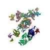
 Movie
Movie Controller


+ Open data

- Basic information
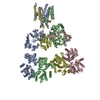
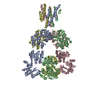
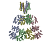
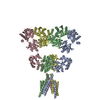
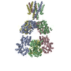
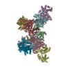
| Entry | Database: PDB / ID: 6l6f | ||||||
|---|---|---|---|---|---|---|---|
| Title | GluK3 receptor complex with UBP301 | ||||||
 Components Components | Glutamate receptor ionotropic, kainate 3 | ||||||
 Keywords Keywords | MEMBRANE PROTEIN | ||||||
| Function / homology |  Function and homology information Function and homology informationPresynaptic function of Kainate receptors / cochlear hair cell ribbon synapse / adenylate cyclase inhibiting G protein-coupled glutamate receptor activity / G protein-coupled glutamate receptor signaling pathway / Activation of Ca-permeable Kainate Receptor / kainate selective glutamate receptor complex / negative regulation of synaptic transmission, glutamatergic / glutamate receptor activity / glutamate receptor signaling pathway / kainate selective glutamate receptor activity ...Presynaptic function of Kainate receptors / cochlear hair cell ribbon synapse / adenylate cyclase inhibiting G protein-coupled glutamate receptor activity / G protein-coupled glutamate receptor signaling pathway / Activation of Ca-permeable Kainate Receptor / kainate selective glutamate receptor complex / negative regulation of synaptic transmission, glutamatergic / glutamate receptor activity / glutamate receptor signaling pathway / kainate selective glutamate receptor activity / glutamate-gated receptor activity / glutamate-gated calcium ion channel activity / dendrite cytoplasm / ligand-gated monoatomic ion channel activity involved in regulation of presynaptic membrane potential / synaptic transmission, glutamatergic / transmitter-gated monoatomic ion channel activity involved in regulation of postsynaptic membrane potential / regulation of membrane potential / postsynaptic density membrane / modulation of chemical synaptic transmission / terminal bouton / presynaptic membrane / chemical synaptic transmission / perikaryon / postsynaptic membrane / axon / dendrite / glutamatergic synapse / plasma membrane Similarity search - Function | ||||||
| Biological species |  | ||||||
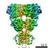
| Method | ELECTRON MICROSCOPY / single particle reconstruction / cryo EM / Resolution: 10.6 Å | ||||||
 Authors Authors | Kumar, J. / Kumari, J. / Burada, A.P. | ||||||
| Funding support |  India, 1items India, 1items
| ||||||
 Citation Citation | Journal: Int J Biol Macromol / Year: 2020 Title: Structural dynamics of the GluK3-kainate receptor neurotransmitter binding domains revealed by cryo-EM. Authors: Jyoti Kumari / Ameya D Bendre / Sumedha Bhosale / Rajesh Vinnakota / Ananth P Burada / Giancarlo Tria / Raimond B G Ravelli / Peter J Peters / Manali Joshi / Janesh Kumar /  Abstract: Kainate receptors belong to the ionotropic glutamate receptor family and play critical roles in the regulation of synaptic networks. The kainate receptor subunit GluK3 has unique functional ...Kainate receptors belong to the ionotropic glutamate receptor family and play critical roles in the regulation of synaptic networks. The kainate receptor subunit GluK3 has unique functional properties and contributes to presynaptic facilitation at the hippocampal mossy fiber synapses along with roles at the post-synapses. To gain structural insights into the unique functional properties and dynamics of GluK3 receptor, we imaged them via electron microscopy in the apo-state and in complex with either agonist kainate or antagonist UBP301. Our analysis of all the GluK3 full-length structures not only provides insights into the receptor transitions between desensitized and closed states but also reveals a "non-classical" conformation of neurotransmitter binding domain in the closed-state distinct from that observed in AMPA and other kainate receptor structures. We show by molecular dynamics simulations that Asp759 influences the stability of the LBD dimers and hence could be responsible for the observed conformational variability and dynamics of the GluK3 via electron microscopy. Lower dimer stability could explain faster desensitization and low agonist sensitivity of GluK3. In overview, our work helps to associate biochemistry and physiology of GluK3 receptors with their structural biology and offers structural insights into the unique functional properties of these atypical receptors. | ||||||
| History |
|
- Structure visualization
| Movie |
Movie viewer |
|---|---|
| Structure viewer | Molecule: MolmilJmol/JSmol |
- Downloads & links
-Download
| PDBx/mmCIF format | 6l6f.cif.gz | 495.6 KB | Display | PDBx/mmCIF format |
|---|---|---|---|---|
| PDB format | pdb6l6f.ent.gz | 408.5 KB | Display | PDB format |
| PDBx/mmJSON format | 6l6f.json.gz | Tree view | PDBx/mmJSON format | |
| Others |  Other downloads Other downloads |
-Validation report
| Arichive directory | https://data.pdbj.org/pub/pdb/validation_reports/l6/6l6fftp://data.pdbj.org/pub/pdb/validation_reports/l6/6l6f | HTTPS FTP |
|---|
-Related structure data
| Related structure data |  0839MC  0790C 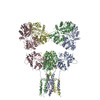 6kzmC M: map data used to model this data C: citing same article ( |
|---|---|
| Similar structure data |

-Links
 PDBj
PDBj

- Assembly
| Deposited unit | 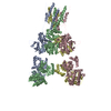
|
|---|---|
| 1 |
|
-Components
| #1: Protein | Mass: 93984.922 Da / Num. of mol.: 4 / Mutation: T117C, T336C, V578C Source method: isolated from a genetically manipulated source Source: (gene. exp.)  Homo sapiens (human) / References: UniProt: G3V9I2, UniProt: P42264*PLUS Homo sapiens (human) / References: UniProt: G3V9I2, UniProt: P42264*PLUSHas protein modification | Y | |
|---|
-Experimental details
-Experiment
| Experiment | Method: ELECTRON MICROSCOPY |
|---|---|
| EM experiment | Aggregation state: PARTICLE / 3D reconstruction method: single particle reconstruction |
- Sample preparation
| Component | Name: GluK3 complex with agonist Kainate / Type: COMPLEX / Entity ID: all / Source: RECOMBINANT |
|---|---|
| Molecular weight | Experimental value: YES |
| Source (natural) | Organism: |
| Source (recombinant) | Organism: Homo sapiens (human) / Cell: HEK293 GnTi- / Plasmid: pEGBacMam |
| Buffer solution | pH: 8 / Details: 20 mM Tris pH 8.0, 150 mM NaCl |
| Buffer component | Conc.: 1.7 1.7 / Name: Sodium Chloride / Formula: NaCl |
| Specimen | Conc.: 1.7 mg/ml / Embedding applied: NO / Shadowing applied: NO / Staining applied: NO / Vitrification applied: YES |
| Specimen support | Grid material: GOLD / Grid mesh size: 300 divisions/in. / Grid type: Quantifoil, UltrAuFoil, R1.2/1.3 |
| Vitrification | Cryogen name: ETHANE / Details: Multiple application, blotted for 3 seconds |
- Electron microscopy imaging
| Experimental equipment |  Model: Talos Arctica / Image courtesy: FEI Company |
|---|---|
| Microscopy | Model: FEI TECNAI ARCTICA |
| Electron gun | Electron source:  FIELD EMISSION GUN / Accelerating voltage: 200 kV / Illumination mode: SPOT SCAN FIELD EMISSION GUN / Accelerating voltage: 200 kV / Illumination mode: SPOT SCAN |
| Electron lens | Mode: BRIGHT FIELD |
| Image recording | Electron dose: 16.73 e/Å2 / Film or detector model: FEI FALCON III (4k x 4k) / Num. of real images: 2845 |
- Processing
| Software | Name: PHENIX / Version: 1.16_3549: / Classification: refinement | ||||||||||||||||||||||||||||||||
|---|---|---|---|---|---|---|---|---|---|---|---|---|---|---|---|---|---|---|---|---|---|---|---|---|---|---|---|---|---|---|---|---|---|
| EM software |
| ||||||||||||||||||||||||||||||||
| CTF correction | Type: PHASE FLIPPING AND AMPLITUDE CORRECTION | ||||||||||||||||||||||||||||||||
| Particle selection | Num. of particles selected: 46106 | ||||||||||||||||||||||||||||||||
| Symmetry | Point symmetry: C1 (asymmetric) | ||||||||||||||||||||||||||||||||
| 3D reconstruction | Resolution: 10.6 Å / Resolution method: FSC 0.143 CUT-OFF / Num. of particles: 27997 / Symmetry type: POINT | ||||||||||||||||||||||||||||||||
| Atomic model building | Protocol: RIGID BODY FIT | ||||||||||||||||||||||||||||||||
| Atomic model building | PDB-ID: 6JFY Accession code: 6JFY / Source name: PDB / Type: experimental model | ||||||||||||||||||||||||||||||||
| Refine LS restraints |
|

