 Movie
Movie Controller

[English] 日本語
 Yorodumi
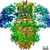
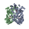
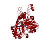
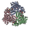
- PDB-6iww: Cryo-EM structure of the S. typhimurium oxaloacetate decarboxylas... -
+ Open data

- Basic information
| Entry | Database: PDB / ID: 6iww | ||||||
|---|---|---|---|---|---|---|---|
| Title | Cryo-EM structure of the S. typhimurium oxaloacetate decarboxylase beta-gamma sub-complex | ||||||
 Components Components |
| ||||||
 Keywords Keywords | MEMBRANE PROTEIN / sodium pump / decarboxylase sodium pump / biotin-dependent decarboxylase | ||||||
| Function / homology |  Function and homology information Function and homology informationoxaloacetate decarboxylase (Na+ extruding) / decarboxylation-driven active transmembrane transporter activity / sodium ion transmembrane transporter activity / oxaloacetate decarboxylase activity / sodium ion export across plasma membrane / sodium ion transport / lyase activity / plasma membrane Similarity search - Function | ||||||
| Biological species |  Salmonella enterica subsp. enterica serovar Typhimurium (bacteria) Salmonella enterica subsp. enterica serovar Typhimurium (bacteria) | ||||||
| Method | ELECTRON MICROSCOPY / single particle reconstruction / cryo EM / Resolution: 3.9 Å | ||||||
 Authors Authors | Xu, X. / Shi, H. / Zhang, X. / Xiang, S. | ||||||
| Funding support |  China, 1items China, 1items
| ||||||
 Citation Citation | Journal: Elife / Year: 2020 Title: Structural insights into sodium transport by the oxaloacetate decarboxylase sodium pump. Authors: Xin Xu / Huigang Shi / Xiaowen Gong / Pu Chen / Ying Gao / Xinzheng Zhang / Song Xiang / Abstract: The oxaloacetate decarboxylase sodium pump (OAD) is a unique primary-active transporter that utilizes the free energy derived from oxaloacetate decarboxylation for sodium transport across the cell ...The oxaloacetate decarboxylase sodium pump (OAD) is a unique primary-active transporter that utilizes the free energy derived from oxaloacetate decarboxylation for sodium transport across the cell membrane. It is composed of 3 subunits: the α subunit catalyzes carboxyl-transfer from oxaloacetate to biotin, the membrane integrated β subunit catalyzes the subsequent carboxyl-biotin decarboxylation and the coupled sodium transport, the γ subunit interacts with the α and β subunits and stabilizes the OAD complex. We present here structure of the OAD βγ sub-complex. The structure revealed that the β and γ subunits form a βγ hetero-hexamer with extensive interactions between the subunits and shed light on the OAD holo-enzyme assembly. Structure-guided functional studies provided insights into the sodium binding sites in the β subunit and the coupling between carboxyl-biotin decarboxylation and sodium transport by the OAD β subunit. | ||||||
| History |
|
- Structure visualization
| Movie |
Movie viewer |
|---|---|
| Structure viewer | Molecule: MolmilJmol/JSmol |
- Downloads & links
-Download
| PDBx/mmCIF format | 6iww.cif.gz | 231.2 KB | Display | PDBx/mmCIF format |
|---|---|---|---|---|
| PDB format | pdb6iww.ent.gz | 186.3 KB | Display | PDB format |
| PDBx/mmJSON format | 6iww.json.gz | Tree view | PDBx/mmJSON format | |
| Others |  Other downloads Other downloads |
-Validation report
| Summary document | 6iww_validation.pdf.gz | 1.1 MB | Display | wwPDB validaton report |
|---|---|---|---|---|
| Full document | 6iww_full_validation.pdf.gz | 1.1 MB | Display | |
| Data in XML | 6iww_validation.xml.gz | 40.4 KB | Display | |
| Data in CIF | 6iww_validation.cif.gz | 61.3 KB | Display | |
| Arichive directory | https://data.pdbj.org/pub/pdb/validation_reports/iw/6iwwftp://data.pdbj.org/pub/pdb/validation_reports/iw/6iww | HTTPS FTP |
-Related structure data
| Related structure data | 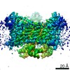 9743MC  6ivaC M: map data used to model this data C: citing same article ( |
|---|---|
| Similar structure data |


-Links
 PDBj
PDBj- Assembly
| Deposited unit | 
|
|---|---|
| 1 |
|
-Components
| #1: Protein | Mass: 44928.801 Da / Num. of mol.: 3 Source method: isolated from a genetically manipulated source Source: (gene. exp.) Salmonella enterica subsp. enterica serovar Typhimurium (bacteria)Strain: enterica serovar Typhimurium / Production host: References: UniProt: A0A0F7JAV8, UniProt: Q8ZRY4*PLUS, oxaloacetate decarboxylase (Na+ extruding) #2: Protein | Mass: 11137.029 Da / Num. of mol.: 3 Source method: isolated from a genetically manipulated source Source: (gene. exp.) Salmonella enterica subsp. enterica serovar Typhimurium (bacteria)Strain: enterica serovar Typhimurium / Production host: References: UniProt: A0A0F7JC72, UniProt: Q03032*PLUS, oxaloacetate decarboxylase (Na+ extruding) #3: Sugar |   Type: D-saccharide / Mass: 510.615 Da / Num. of mol.: 3 / Source method: obtained synthetically / Formula: C24H46O11 / Comment: detergent*YM Type: D-saccharide / Mass: 510.615 Da / Num. of mol.: 3 / Source method: obtained synthetically / Formula: C24H46O11 / Comment: detergent*YMSequence details | Authors state that this conflict may be a natural occurring mutation that happen to exist in the ...Authors state that this conflict may be a natural occurring mutation that happen to exist in the genomic DNA they used. | |
|---|
-Experimental details
-Experiment
| Experiment | Method: ELECTRON MICROSCOPY |
|---|---|
| EM experiment | Aggregation state: PARTICLE / 3D reconstruction method: single particle reconstruction |
- Sample preparation
| Component | Name: The oxaloacetate decarboxylase beta-gamma sub-complex / Type: COMPLEX / Entity ID: #2 / Source: RECOMBINANT |
|---|---|
| Molecular weight | Experimental value: NO |
| Source (natural) | Organism: Salmonella enterica subsp. enterica serovar Typhimurium (bacteria) Strain: enterica serovar Typhimurium |
| Source (recombinant) | Organism: |
| Buffer solution | pH: 7.5 |
| Specimen | Conc.: 8 mg/ml / Embedding applied: NO / Shadowing applied: NO / Staining applied: NO / Vitrification applied: YES / Details: This sample was monodisperse |
| Specimen support | Grid material: COPPER / Grid type: Quantifoil R1.2/1.3 |
| Vitrification | Cryogen name: ETHANE |
- Electron microscopy imaging
| Experimental equipment |  Model: Titan Krios / Image courtesy: FEI Company |
|---|---|
| Microscopy | Model: FEI TITAN KRIOS |
| Electron gun | Electron source:  FIELD EMISSION GUN / Accelerating voltage: 300 kV / Illumination mode: FLOOD BEAM FIELD EMISSION GUN / Accelerating voltage: 300 kV / Illumination mode: FLOOD BEAM |
| Electron lens | Mode: BRIGHT FIELD / Calibrated defocus min: 1500 nm / Calibrated defocus max: 3500 nm |
| Specimen holder | Cryogen: NITROGEN / Specimen holder model: FEI TITAN KRIOS AUTOGRID HOLDER |
| Image recording | Electron dose: 50 e/Å2 / Detector mode: SUPER-RESOLUTION / Film or detector model: GATAN K2 SUMMIT (4k x 4k) / Num. of grids imaged: 1 / Num. of real images: 821 |
| Image scans | Movie frames/image: 32 / Used frames/image: 1-50 |
- Processing
| Software | Name: PHENIX / Version: 1.13_2998: / Classification: refinement | ||||||||||||||||||||||||||||||||
|---|---|---|---|---|---|---|---|---|---|---|---|---|---|---|---|---|---|---|---|---|---|---|---|---|---|---|---|---|---|---|---|---|---|
| EM software |
| ||||||||||||||||||||||||||||||||
| CTF correction | Type: PHASE FLIPPING ONLY | ||||||||||||||||||||||||||||||||
| Symmetry | Point symmetry: C3 (3 fold cyclic) | ||||||||||||||||||||||||||||||||
| 3D reconstruction | Resolution: 3.9 Å / Resolution method: FSC 0.143 CUT-OFF / Num. of particles: 75699 / Algorithm: FOURIER SPACE / Num. of class averages: 1 / Symmetry type: POINT |
