[English] 日本語
 Yorodumi
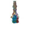
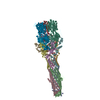
- PDB-6iol: Cryo-EM structure of multidrug efflux pump MexAB-OprM (60 degree ... -
+ Open data
- Basic information
| Entry | Database: PDB / ID: 6iol | ||||||||||||||||||||||||||||||||||||||||||||||||||||||
|---|---|---|---|---|---|---|---|---|---|---|---|---|---|---|---|---|---|---|---|---|---|---|---|---|---|---|---|---|---|---|---|---|---|---|---|---|---|---|---|---|---|---|---|---|---|---|---|---|---|---|---|---|---|---|---|
| Title | Cryo-EM structure of multidrug efflux pump MexAB-OprM (60 degree state) | ||||||||||||||||||||||||||||||||||||||||||||||||||||||
 Components Components |
| ||||||||||||||||||||||||||||||||||||||||||||||||||||||
 Keywords Keywords | MEMBRANE PROTEIN / Multidrug resistance / efflux / complex | ||||||||||||||||||||||||||||||||||||||||||||||||||||||
| Function / homology |  Function and homology information Function and homology informationxenobiotic detoxification by transmembrane export across the cell outer membrane / efflux pump complex / efflux transmembrane transporter activity / xenobiotic transmembrane transporter activity / transmembrane transporter activity / cell outer membrane / protein homooligomerization / transmembrane transport / response to antibiotic / identical protein binding ...xenobiotic detoxification by transmembrane export across the cell outer membrane / efflux pump complex / efflux transmembrane transporter activity / xenobiotic transmembrane transporter activity / transmembrane transporter activity / cell outer membrane / protein homooligomerization / transmembrane transport / response to antibiotic / identical protein binding / membrane / plasma membrane Similarity search - Function | ||||||||||||||||||||||||||||||||||||||||||||||||||||||
| Biological species |   Pseudomonas aeruginosa PAO1 (bacteria) Pseudomonas aeruginosa PAO1 (bacteria) | ||||||||||||||||||||||||||||||||||||||||||||||||||||||
| Method | ELECTRON MICROSCOPY / single particle reconstruction / cryo EM / Resolution: 3.76 Å | ||||||||||||||||||||||||||||||||||||||||||||||||||||||
 Authors Authors | Tsutsumi, K. / Yonehara, R. / Nakagawa, A. / Yamashita, E. | ||||||||||||||||||||||||||||||||||||||||||||||||||||||
| Funding support |  Japan, 3items Japan, 3items
| ||||||||||||||||||||||||||||||||||||||||||||||||||||||
 Citation Citation |  Journal: Nat Commun / Year: 2019 Journal: Nat Commun / Year: 2019Title: Structures of the wild-type MexAB-OprM tripartite pump reveal its complex formation and drug efflux mechanism. Authors: Kenta Tsutsumi / Ryo Yonehara / Etsuko Ishizaka-Ikeda / Naoyuki Miyazaki / Shintaro Maeda / Kenji Iwasaki / Atsushi Nakagawa / Eiki Yamashita /   Abstract: In Pseudomonas aeruginosa, MexAB-OprM plays a central role in multidrug resistance by ejecting various drug compounds, which is one of the causes of serious nosocomial infections. Although the ...In Pseudomonas aeruginosa, MexAB-OprM plays a central role in multidrug resistance by ejecting various drug compounds, which is one of the causes of serious nosocomial infections. Although the structures of the components of MexAB-OprM have been solved individually by X-ray crystallography, no structural information for fully assembled pumps from P. aeruginosa were previously available. In this study, we present the structure of wild-type MexAB-OprM in the presence or absence of drugs at near-atomic resolution. The structure reveals that OprM does not interact with MexB directly, and that it opens its periplasmic gate by forming a complex. Furthermore, we confirm the residues essential for complex formation and observed a movement of the drug entrance gate. Based on these results, we propose mechanisms for complex formation and drug efflux. | ||||||||||||||||||||||||||||||||||||||||||||||||||||||
| History |
|
- Structure visualization
| Movie |
 Movie viewer Movie viewer |
|---|---|
| Structure viewer | Molecule:  Molmil Molmil Jmol/JSmol Jmol/JSmol |
- Downloads & links
- Download
| PDBx/mmCIF format |  6iol.cif.gz 6iol.cif.gz | 1 MB | Display |  PDBx/mmCIF format PDBx/mmCIF format |
|---|---|---|---|---|
| PDB format |  pdb6iol.ent.gz pdb6iol.ent.gz | 878.9 KB | Display |  PDB format PDB format |
| PDBx/mmJSON format |  6iol.json.gz 6iol.json.gz | Tree view |  PDBx/mmJSON format PDBx/mmJSON format | |
| Others |  Other downloads Other downloads |
-Validation report
| Arichive directory |  https://data.pdbj.org/pub/pdb/validation_reports/io/6iol https://data.pdbj.org/pub/pdb/validation_reports/io/6iol ftp://data.pdbj.org/pub/pdb/validation_reports/io/6iol ftp://data.pdbj.org/pub/pdb/validation_reports/io/6iol | HTTPS FTP |
|---|
-Related structure data
| Related structure data |  9696MC  9695C  6iokC M: map data used to model this data C: citing same article ( |
|---|---|
| Similar structure data | |
| EM raw data |  EMPIAR-10292 (Title: Cryo-EM structure of multidrug efflux pump MexAB-OprM EMPIAR-10292 (Title: Cryo-EM structure of multidrug efflux pump MexAB-OprMData size: 8.5 TB Data #1: Raw multiframe micrographs [micrographs - multiframe]) |
- Links
Links
- Assembly
| Deposited unit | 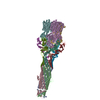
|
|---|---|
| 1 |
|
- Components
| #1: Protein | Mass: 38789.695 Da / Num. of mol.: 6 / Fragment: UNP residues 25-383 Source method: isolated from a genetically manipulated source Source: (gene. exp.)   #2: Protein | Mass: 51737.605 Da / Num. of mol.: 3 / Fragment: UNP residues 18-485 Source method: isolated from a genetically manipulated source Source: (gene. exp.)  Pseudomonas aeruginosa PAO1 (bacteria) / Strain: PAO1 / Gene: oprM / Production host: Pseudomonas aeruginosa PAO1 (bacteria) / Strain: PAO1 / Gene: oprM / Production host:  #3: Protein | Mass: 113948.273 Da / Num. of mol.: 3 Source method: isolated from a genetically manipulated source Source: (gene. exp.)  Pseudomonas aeruginosa PAO1 (bacteria) / Strain: PAO1 / Gene: mexB / Production host: Pseudomonas aeruginosa PAO1 (bacteria) / Strain: PAO1 / Gene: mexB / Production host:  Has protein modification | N | |
|---|
-Experimental details
-Experiment
| Experiment | Method: ELECTRON MICROSCOPY |
|---|---|
| EM experiment | Aggregation state: PARTICLE / 3D reconstruction method: single particle reconstruction |
- Sample preparation
| Component | Name: membrane protein complex / Type: COMPLEX / Entity ID: #1 / Source: RECOMBINANT |
|---|---|
| Molecular weight | Value: 0.73 MDa / Experimental value: NO |
| Source (natural) | Organism:  |
| Source (recombinant) | Organism:  |
| Buffer solution | pH: 7.5 |
| Buffer component | Conc.: 50 mM / Name: HEPES / Formula: C8H18N2O4S |
| Specimen | Conc.: 9.1 mg/ml / Embedding applied: NO / Shadowing applied: NO / Staining applied: NO / Vitrification applied: YES |
| Vitrification | Instrument: FEI VITROBOT MARK IV / Cryogen name: ETHANE / Chamber temperature: 277 K |
- Electron microscopy imaging
| Experimental equipment |  Model: Titan Krios / Image courtesy: FEI Company |
|---|---|
| Microscopy | Model: FEI TITAN KRIOS |
| Electron gun | Electron source:  FIELD EMISSION GUN / Accelerating voltage: 300 kV / Illumination mode: FLOOD BEAM FIELD EMISSION GUN / Accelerating voltage: 300 kV / Illumination mode: FLOOD BEAM |
| Electron lens | Mode: BRIGHT FIELD / Nominal magnification: 75000 X / Nominal defocus max: 3000 nm / Nominal defocus min: 1250 nm / Cs: 0.1 mm / C2 aperture diameter: 100 µm / Alignment procedure: ZEMLIN TABLEAU |
| Specimen holder | Cryogen: NITROGEN / Specimen holder model: FEI TITAN KRIOS AUTOGRID HOLDER / Temperature (max): 95 K / Temperature (min): 90 K |
| Image recording | Average exposure time: 2 sec. / Electron dose: 40 e/Å2 / Detector mode: INTEGRATING / Film or detector model: FEI FALCON II (4k x 4k) / Num. of grids imaged: 1 / Num. of real images: 8722 |
| EM imaging optics | Spherical aberration corrector: CEOS Cs-corrector |
| Image scans | Sampling size: 14 µm / Width: 4080 / Height: 4080 / Movie frames/image: 32 / Used frames/image: 1-30 |
- Processing
| Software | Name: PHENIX / Version: 1.14_3260: / Classification: refinement | ||||||||||||||||||||||||
|---|---|---|---|---|---|---|---|---|---|---|---|---|---|---|---|---|---|---|---|---|---|---|---|---|---|
| EM software |
| ||||||||||||||||||||||||
| CTF correction | Type: PHASE FLIPPING AND AMPLITUDE CORRECTION | ||||||||||||||||||||||||
| Particle selection | Num. of particles selected: 535948 | ||||||||||||||||||||||||
| Symmetry | Point symmetry: C1 (asymmetric) | ||||||||||||||||||||||||
| 3D reconstruction | Resolution: 3.76 Å / Resolution method: FSC 0.143 CUT-OFF / Num. of particles: 42338 / Algorithm: BACK PROJECTION / Symmetry type: POINT | ||||||||||||||||||||||||
| Refine LS restraints |
|
 Movie
Movie Controller


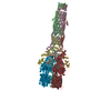
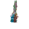
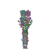


 PDBj
PDBj


