 Movie
Movie Controller

[English] 日本語
 Yorodumi
- PDB-6gsi: Feline Calicivirus Strain F9 bound to a soluble ectodomain fragme... -
+ Open data

- Basic information
| Entry | Database: PDB / ID: 6gsi | |||||||||
|---|---|---|---|---|---|---|---|---|---|---|
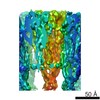
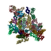
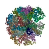
| Title | Feline Calicivirus Strain F9 bound to a soluble ectodomain fragment of feline junctional adhesion molecule A - leading to assembly of a portal structure at a unique three-fold axis. | |||||||||
 Components Components |
| |||||||||
 Keywords Keywords | VIRUS / Capsid / Calicivirus / Vesivirus / Vp1 / portal / Vp2 / junctional-adhesion molecule A | |||||||||
| Function / homology |  Function and homology information Function and homology informationestablishment of endothelial intestinal barrier / regulation of membrane permeability / T=3 icosahedral viral capsid / intestinal absorption / bicellular tight junction / host cell cytoplasm / membrane => GO:0016020 / cell adhesion / plasma membrane Similarity search - Function | |||||||||
| Biological species |   Feline calicivirus strain F9 Feline calicivirus strain F9 | |||||||||
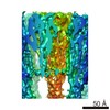
| Method | ELECTRON MICROSCOPY / single particle reconstruction / cryo EM / Resolution: 3.75 Å | |||||||||
 Authors Authors | Conley, M.J. / Bhella, D. | |||||||||
| Funding support |  United Kingdom, 2items United Kingdom, 2items
| |||||||||
 Citation Citation | Journal: Nature / Year: 2019 Title: Calicivirus VP2 forms a portal-like assembly following receptor engagement. Authors: Michaela J Conley / Marion McElwee / Liyana Azmi / Mads Gabrielsen / Olwyn Byron / Ian G Goodfellow / David Bhella / Abstract: To initiate infection, many viruses enter their host cells by triggering endocytosis following receptor engagement. However, the mechanisms by which non-enveloped viruses escape the endosome are ...To initiate infection, many viruses enter their host cells by triggering endocytosis following receptor engagement. However, the mechanisms by which non-enveloped viruses escape the endosome are poorly understood. Here we present near-atomic-resolution cryo-electron microscopy structures for feline calicivirus both undecorated and labelled with a soluble fragment of its cellular receptor, feline junctional adhesion molecule A. We show that VP2, a minor capsid protein encoded by all caliciviruses, forms a large portal-like assembly at a unique three-fold axis of symmetry, following receptor engagement. This assembly-which was not detected in undecorated virions-is formed of twelve copies of VP2, arranged with their hydrophobic N termini pointing away from the virion surface. Local rearrangement at the portal site leads to the opening of a pore in the capsid shell. We hypothesize that the portal-like assembly functions as a channel for the delivery of the calicivirus genome, through the endosomal membrane, into the cytoplasm of a host cell, thereby initiating infection. VP2 was previously known to be critical for the production of infectious virus; our findings provide insights into its structure and function that advance our understanding of the Caliciviridae. | |||||||||
| History |
|
- Structure visualization
| Movie |
Movie viewer |
|---|---|
| Structure viewer | Molecule: MolmilJmol/JSmol |
- Downloads & links
-Download
| PDBx/mmCIF format | 6gsi.cif.gz | 1 MB | Display | PDBx/mmCIF format |
|---|---|---|---|---|
| PDB format | pdb6gsi.ent.gz | 889 KB | Display | PDB format |
| PDBx/mmJSON format | 6gsi.json.gz | Tree view | PDBx/mmJSON format | |
| Others |  Other downloads Other downloads |
-Validation report
| Summary document | 6gsi_validation.pdf.gz | 1.1 MB | Display | wwPDB validaton report |
|---|---|---|---|---|
| Full document | 6gsi_full_validation.pdf.gz | 1.1 MB | Display | |
| Data in XML | 6gsi_validation.xml.gz | 95.9 KB | Display | |
| Data in CIF | 6gsi_validation.cif.gz | 147.8 KB | Display | |
| Arichive directory | https://data.pdbj.org/pub/pdb/validation_reports/gs/6gsiftp://data.pdbj.org/pub/pdb/validation_reports/gs/6gsi | HTTPS FTP |
-Related structure data
| Related structure data | 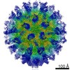 0056MC 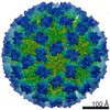 0054C 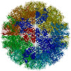 6gshC C: citing same article ( M: map data used to model this data |
|---|---|
| Similar structure data | |
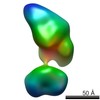
| EM raw data | EMPIAR-10193 (Title: Calicivirus VP2 forms a portal to mediate endosome escape Data size: 867.0 Data #1: Motion corrected micrographs of feline calicivirus strain F9 decorated with feline junctional adhesion molecule A [micrographs - single frame]) |
-Links
 PDBj
PDBj


- Assembly
| Deposited unit | 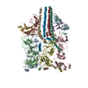
|
|---|---|
| 1 | 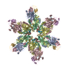
|
-Components
| #1: Protein | Mass: 73346.664 Da / Num. of mol.: 4 / Source method: isolated from a natural source / Source: (natural) Feline calicivirus strain F9 / References: UniProt: P27406#2: Protein | Mass: 22180.650 Da / Num. of mol.: 4 / Fragment: UNP residues 29-230 Source method: isolated from a genetically manipulated source Source: (gene. exp.)  Cricetulus griseus (Chinese hamster) / References: UniProt: Q2WGK2 Cricetulus griseus (Chinese hamster) / References: UniProt: Q2WGK2#3: Protein | Mass: 12209.838 Da / Num. of mol.: 4 / Source method: isolated from a natural source / Source: (natural) Feline calicivirus strain F9 / References: UniProt: P28711#4: Chemical | ChemComp-K /   Mass: 39.098 Da / Num. of mol.: 4 / Source method: obtained synthetically / Formula: K Mass: 39.098 Da / Num. of mol.: 4 / Source method: obtained synthetically / Formula: K |
|---|
-Experimental details
-Experiment
| Experiment | Method: ELECTRON MICROSCOPY |
|---|---|
| EM experiment | Aggregation state: PARTICLE / 3D reconstruction method: single particle reconstruction |
- Sample preparation
| Component |
| |||||||||||||||||||||||||||||||||||
|---|---|---|---|---|---|---|---|---|---|---|---|---|---|---|---|---|---|---|---|---|---|---|---|---|---|---|---|---|---|---|---|---|---|---|---|---|
| Molecular weight | Experimental value: NO | |||||||||||||||||||||||||||||||||||
| Source (natural) |
| |||||||||||||||||||||||||||||||||||
| Source (recombinant) | Organism: Cricetulus griseus (Chinese hamster) / Cell: CHO cells | |||||||||||||||||||||||||||||||||||
| Details of virus |
| |||||||||||||||||||||||||||||||||||
| Natural host |
| |||||||||||||||||||||||||||||||||||
| Virus shell |
| |||||||||||||||||||||||||||||||||||
| Buffer solution | pH: 7.2 / Details: Phosphate buffered saline | |||||||||||||||||||||||||||||||||||
| Specimen | Conc.: 1 mg/ml / Embedding applied: NO / Shadowing applied: NO / Staining applied: NO / Vitrification applied: YES Details: Purified virions were incubated in the presence of purified ectodomain fragments. | |||||||||||||||||||||||||||||||||||
| Specimen support | Grid material: COPPER / Grid mesh size: 400 divisions/in. / Grid type: C-flat-2/2 | |||||||||||||||||||||||||||||||||||
| Vitrification | Instrument: FEI VITROBOT MARK IV / Cryogen name: ETHANE / Humidity: 100 % / Chamber temperature: 277 K |
- Electron microscopy imaging
| Experimental equipment |  Model: Titan Krios / Image courtesy: FEI Company |
|---|---|
| Microscopy | Model: FEI TITAN KRIOS |
| Electron gun | Electron source:  FIELD EMISSION GUN / Accelerating voltage: 300 kV / Illumination mode: FLOOD BEAM FIELD EMISSION GUN / Accelerating voltage: 300 kV / Illumination mode: FLOOD BEAM |
| Electron lens | Mode: BRIGHT FIELD / Nominal magnification: 75000 X / Cs: 2.7 mm |
| Specimen holder | Cryogen: NITROGEN / Specimen holder model: FEI TITAN KRIOS AUTOGRID HOLDER |
| Image recording | Electron dose: 63 e/Å2 / Detector mode: INTEGRATING / Film or detector model: FEI FALCON III (4k x 4k) / Num. of grids imaged: 1 / Num. of real images: 13865 Details: Each micrograph was recorded as a movie of 50 individual fractions with a total dose of 63 e/angstrom squared |
- Processing
| Software | Name: PHENIX / Version: 1.13_2998: / Classification: refinement | ||||||||||||||||||||||||||||
|---|---|---|---|---|---|---|---|---|---|---|---|---|---|---|---|---|---|---|---|---|---|---|---|---|---|---|---|---|---|
| EM software |
| ||||||||||||||||||||||||||||
| Image processing | Details: Images were motion-corrected using motioncor2 Defocus estimation was performed using GCTF | ||||||||||||||||||||||||||||
| CTF correction | Details: CTF correction was implemented through Relion / Type: PHASE FLIPPING AND AMPLITUDE CORRECTION | ||||||||||||||||||||||||||||
| Particle selection | Num. of particles selected: 129884 / Details: Autopicking in Relion | ||||||||||||||||||||||||||||
| Symmetry | Point symmetry: C1 (asymmetric) | ||||||||||||||||||||||||||||
| 3D reconstruction | Resolution: 3.75 Å / Resolution method: FSC 0.143 CUT-OFF / Num. of particles: 58510 Details: Origins and orientations were originally assigned by 3D auto refinement with full icosahedral symmetry. Random group assignment was carried through the focussed classification process and ...Details: Origins and orientations were originally assigned by 3D auto refinement with full icosahedral symmetry. Random group assignment was carried through the focussed classification process and used to divide the data into two roughly even halves that had been initially refined independently. Symmetry type: POINT |
