 Movie
Movie Controller

[English] 日本語
 Yorodumi
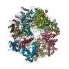
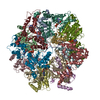
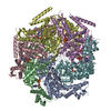
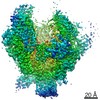
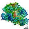
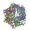
- PDB-6ef0: Yeast 26S proteasome bound to ubiquitinated substrate (1D* motor ... -
+ Open data

- Basic information
| Entry | Database: PDB / ID: 6ef0 | |||||||||
|---|---|---|---|---|---|---|---|---|---|---|
| Title | Yeast 26S proteasome bound to ubiquitinated substrate (1D* motor state) | |||||||||
 Components Components |
| |||||||||
 Keywords Keywords | MOTOR PROTEIN / 26S Proteasome / ATPase / AAA+ / Protease / Ubiquitin | |||||||||
| Function / homology |  Function and homology information Function and homology informationmitotic cell cycle phase transition / proteasome regulatory particle assembly / proteasome-activating activity / protein-containing complex localization / proteasome regulatory particle, base subcomplex / ER-Phagosome pathway / Antigen processing: Ub, ATP-independent proteasomal degradation / cyclin-dependent protein serine/threonine kinase regulator activity / proteasome core complex assembly / Proteasome assembly ...mitotic cell cycle phase transition / proteasome regulatory particle assembly / proteasome-activating activity / protein-containing complex localization / proteasome regulatory particle, base subcomplex / ER-Phagosome pathway / Antigen processing: Ub, ATP-independent proteasomal degradation / cyclin-dependent protein serine/threonine kinase regulator activity / proteasome core complex assembly / Proteasome assembly / Cross-presentation of soluble exogenous antigens (endosomes) / TNFR2 non-canonical NF-kB pathway / nuclear outer membrane-endoplasmic reticulum membrane network / nonfunctional rRNA decay / Regulation of PTEN stability and activity / CDK-mediated phosphorylation and removal of Cdc6 / FBXL7 down-regulates AURKA during mitotic entry and in early mitosis / KEAP1-NFE2L2 pathway / Neddylation / Orc1 removal from chromatin / peptide catabolic process / Ubiquitin-Mediated Degradation of Phosphorylated Cdc25A / MAPK6/MAPK4 signaling / Antigen processing: Ubiquitination & Proteasome degradation / proteasomal ubiquitin-independent protein catabolic process / positive regulation of RNA polymerase II transcription preinitiation complex assembly / Ub-specific processing proteases / proteasome storage granule / proteasome core complex, alpha-subunit complex / Neutrophil degranulation / ERAD pathway / proteasome complex / nucleotide-excision repair / positive regulation of transcription elongation by RNA polymerase II / positive regulation of protein catabolic process / ubiquitin-dependent protein catabolic process / proteasome-mediated ubiquitin-dependent protein catabolic process / chromatin remodeling / protein domain specific binding / cell division / mRNA binding / ubiquitin protein ligase binding / ATP hydrolysis activity / mitochondrion / ATP binding / identical protein binding / nucleus / cytosol / cytoplasm Similarity search - Function | |||||||||
| Biological species |   Homo sapiens (human) Homo sapiens (human) | |||||||||
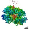
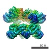
| Method | ELECTRON MICROSCOPY / single particle reconstruction / cryo EM / Resolution: 4.43 Å | |||||||||
 Authors Authors | de la Pena, A.H. / Goodall, E.A. / Gates, S.N. / Lander, G.C. / Martin, A. | |||||||||
| Funding support |  United States, 2items United States, 2items
| |||||||||
 Citation Citation | Journal: Science / Year: 2018 Title: Substrate-engaged 26 proteasome structures reveal mechanisms for ATP-hydrolysis-driven translocation. Authors: Andres H de la Peña / Ellen A Goodall / Stephanie N Gates / Gabriel C Lander / Andreas Martin / Abstract: The 26 proteasome is the primary eukaryotic degradation machine and thus is critically involved in numerous cellular processes. The heterohexameric adenosine triphosphatase (ATPase) motor of the ...The 26 proteasome is the primary eukaryotic degradation machine and thus is critically involved in numerous cellular processes. The heterohexameric adenosine triphosphatase (ATPase) motor of the proteasome unfolds and translocates targeted protein substrates into the open gate of a proteolytic core while a proteasomal deubiquitinase concomitantly removes substrate-attached ubiquitin chains. However, the mechanisms by which ATP hydrolysis drives the conformational changes responsible for these processes have remained elusive. Here we present the cryo-electron microscopy structures of four distinct conformational states of the actively ATP-hydrolyzing, substrate-engaged 26 proteasome. These structures reveal how mechanical substrate translocation accelerates deubiquitination and how ATP-binding, -hydrolysis, and phosphate-release events are coordinated within the AAA+ (ATPases associated with diverse cellular activities) motor to induce conformational changes and propel the substrate through the central pore. | |||||||||
| History |
|
- Structure visualization
| Movie |
Movie viewer |
|---|---|
| Structure viewer | Molecule: MolmilJmol/JSmol |
- Downloads & links
-Download
| PDBx/mmCIF format | 6ef0.cif.gz | 571 KB | Display | PDBx/mmCIF format |
|---|---|---|---|---|
| PDB format | pdb6ef0.ent.gz | 451.7 KB | Display | PDB format |
| PDBx/mmJSON format | 6ef0.json.gz | Tree view | PDBx/mmJSON format | |
| Others |  Other downloads Other downloads |
-Validation report
| Arichive directory | https://data.pdbj.org/pub/pdb/validation_reports/ef/6ef0ftp://data.pdbj.org/pub/pdb/validation_reports/ef/6ef0 | HTTPS FTP |
|---|
-Related structure data
| Related structure data | 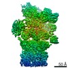 9042MC 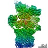 9043C 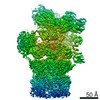 9044C 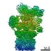 9045C 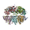 6ef1C 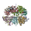 6ef2C 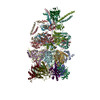 6ef3C M: map data used to model this data C: citing same article ( |
|---|---|
| Similar structure data |
-Links
 PDBj
PDBj


- Assembly
| Deposited unit | 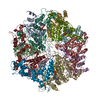
|
|---|---|
| 1 |
|
-Components
-Proteasome subunit alpha type- ... , 6 types, 6 molecules ABCDEF
| #1: Protein | Mass: 26801.549 Da / Num. of mol.: 1 Source method: isolated from a genetically manipulated source Source: (gene. exp.) Strain: ATCC 204508 / S288c / Gene: SCL1, PRC2, PRS2, YGL011C / Production host: References: UniProt: P21243, proteasome endopeptidase complex |
|---|---|
| #2: Protein | Mass: 27191.828 Da / Num. of mol.: 1 Source method: isolated from a genetically manipulated source Source: (gene. exp.) Strain: ATCC 204508 / S288c / Gene: PRE8, PRS4, YML092C / Production host: References: UniProt: P23639, proteasome endopeptidase complex |
| #3: Protein | Mass: 27080.506 Da / Num. of mol.: 1 Source method: isolated from a genetically manipulated source Source: (gene. exp.) Strain: ATCC 204508 / S288c / Gene: PRE9, PRS5, YGR135W / Production host: References: UniProt: P23638, proteasome endopeptidase complex |
| #4: Protein | Mass: 26993.475 Da / Num. of mol.: 1 Source method: isolated from a genetically manipulated source Source: (gene. exp.) Strain: ATCC 204508 / S288c / Gene: PRE6, YOL038W / Production host: References: UniProt: P40303, proteasome endopeptidase complex |
| #5: Protein | Mass: 27444.869 Da / Num. of mol.: 1 Source method: isolated from a genetically manipulated source Source: (gene. exp.) Strain: ATCC 204508 / S288c / Gene: PUP2, DOA5, YGR253C, G9155 / Production host: References: UniProt: P32379, proteasome endopeptidase complex |
| #6: Protein | Mass: 25634.000 Da / Num. of mol.: 1 Source method: isolated from a genetically manipulated source Source: (gene. exp.) Strain: ATCC 204508 / S288c / Gene: PRE5, YMR314W, YM9924.06 / Production host: References: UniProt: P40302, proteasome endopeptidase complex |
-Protein , 2 types, 2 molecules GL
| #7: Protein | Mass: 27092.719 Da / Num. of mol.: 1 Source method: isolated from a genetically manipulated source Source: (gene. exp.) Strain: ATCC 204508 / S288c / Gene: PRE10, PRC1, PRS1, YOR362C, O6650 / Production host: References: UniProt: P21242, proteasome endopeptidase complex |
|---|---|
| #12: Protein | Mass: 30386.865 Da / Num. of mol.: 1 Source method: isolated from a genetically manipulated source Source: (gene. exp.) Strain: ATCC 204508 / S288c / Gene: RPT4, CRL13, PCS1, SUG2, YOR259C / Production host: |
-26S proteasome regulatory subunit ... , 5 types, 5 molecules HIJKM
| #8: Protein | Mass: 28597.023 Da / Num. of mol.: 1 Source method: isolated from a genetically manipulated source Source: (gene. exp.) Strain: ATCC 204508 / S288c / Gene: RPT1, CIM5, YTA3, YKL145W / Production host: |
|---|---|
| #9: Protein | Mass: 30109.572 Da / Num. of mol.: 1 Source method: isolated from a genetically manipulated source Source: (gene. exp.) Strain: ATCC 204508 / S288c / Gene: RPT2, YHS4, YTA5, YDL007W, D2920 / Production host: |
| #10: Protein | Mass: 30522.594 Da / Num. of mol.: 1 Source method: isolated from a genetically manipulated source Source: (gene. exp.) Strain: ATCC 204508 / S288c / Gene: RPT6, CIM3, CRL3, SUG1, TBPY, TBY1, YGL048C / Production host: |
| #11: Protein | Mass: 30387.693 Da / Num. of mol.: 1 Source method: isolated from a genetically manipulated source Source: (gene. exp.) Strain: ATCC 204508 / S288c / Gene: RPT3, YNT1, YTA2, YDR394W, D9509.14 / Production host: |
| #13: Protein | Mass: 28497.504 Da / Num. of mol.: 1 Source method: isolated from a genetically manipulated source Source: (gene. exp.) Strain: ATCC 204508 / S288c / Gene: RPT5, YTA1, YOR117W, O3258, YOR3258W / Production host: |
-Protein/peptide , 1 types, 1 molecules s
| #14: Protein/peptide | Mass: 1130.277 Da / Num. of mol.: 1 Source method: isolated from a genetically manipulated source Source: (gene. exp.) Homo sapiens (human) / Production host:  |
|---|
-Non-polymers , 2 types, 6 molecules 

| #15: Chemical |  Mass: 427.201 Da / Num. of mol.: 3 / Source method: obtained synthetically / Formula: C10H15N5O10P2 / Comment: ADP, energy-carrying molecule*YM Mass: 427.201 Da / Num. of mol.: 3 / Source method: obtained synthetically / Formula: C10H15N5O10P2 / Comment: ADP, energy-carrying molecule*YM#16: Chemical |  Mass: 507.181 Da / Num. of mol.: 3 / Source method: obtained synthetically / Formula: C10H16N5O13P3 / Comment: ATP, energy-carrying molecule*YM Mass: 507.181 Da / Num. of mol.: 3 / Source method: obtained synthetically / Formula: C10H16N5O13P3 / Comment: ATP, energy-carrying molecule*YM |
|---|
-Experimental details
-Experiment
| Experiment | Method: ELECTRON MICROSCOPY |
|---|---|
| EM experiment | Aggregation state: PARTICLE / 3D reconstruction method: single particle reconstruction |
- Sample preparation
| Component |
| ||||||||||||||||||||||||||||||||||||
|---|---|---|---|---|---|---|---|---|---|---|---|---|---|---|---|---|---|---|---|---|---|---|---|---|---|---|---|---|---|---|---|---|---|---|---|---|---|
| Source (natural) |
| ||||||||||||||||||||||||||||||||||||
| Source (recombinant) |
| ||||||||||||||||||||||||||||||||||||
| Buffer solution | pH: 7.6 | ||||||||||||||||||||||||||||||||||||
| Buffer component |
| ||||||||||||||||||||||||||||||||||||
| Specimen | Conc.: 25 mg/ml / Embedding applied: NO / Shadowing applied: NO / Staining applied: NO / Vitrification applied: YES Details: 26S proteasomes were diluted to a concentration of 20 micromolar in a buffer with an ATP regeneration system, and 6 mM ortho-phenanthroline. This solution was mixed with an equal volume of ...Details: 26S proteasomes were diluted to a concentration of 20 micromolar in a buffer with an ATP regeneration system, and 6 mM ortho-phenanthroline. This solution was mixed with an equal volume of 50 micromolar ubiquitinated model substrate | ||||||||||||||||||||||||||||||||||||
| Specimen support | Grid material: COPPER / Grid mesh size: 400 divisions/in. / Grid type: Quantifoil R2/2 | ||||||||||||||||||||||||||||||||||||
| Vitrification | Instrument: HOMEMADE PLUNGER / Cryogen name: ETHANE / Humidity: 90 % / Chamber temperature: 277 K Details: specimens were manually blotted with Whatman #1 filter paper |
- Electron microscopy imaging
| Experimental equipment |  Model: Titan Krios / Image courtesy: FEI Company |
|---|---|
| Microscopy | Model: FEI TITAN KRIOS / Details: images were acquired in nanoprobe mode |
| Electron gun | Electron source:  FIELD EMISSION GUN / Accelerating voltage: 300 kV / Illumination mode: FLOOD BEAM FIELD EMISSION GUN / Accelerating voltage: 300 kV / Illumination mode: FLOOD BEAM |
| Electron lens | Mode: BRIGHT FIELD / Nominal magnification: 29000 X / Nominal defocus max: -2500 nm / Nominal defocus min: -1000 nm / Calibrated defocus min: -1500 nm / Calibrated defocus max: -3000 nm / Cs: 2.7 mm / C2 aperture diameter: 70 µm / Alignment procedure: COMA FREE |
| Specimen holder | Cryogen: NITROGEN / Specimen holder model: FEI TITAN KRIOS AUTOGRID HOLDER |
| Image recording | Average exposure time: 6.25 sec. / Electron dose: 50 e/Å2 / Detector mode: COUNTING / Film or detector model: GATAN K2 SUMMIT (4k x 4k) / Num. of grids imaged: 1 / Num. of real images: 11656 |
| Image scans | Sampling size: 5 µm / Width: 3710 / Height: 3838 / Movie frames/image: 25 / Used frames/image: 1-25 |
- Processing
| Software | Name: PHENIX / Version: 1.11.1_2580: / Classification: refinement | ||||||||||||||||||||||||||||||||||||||||||||||||
|---|---|---|---|---|---|---|---|---|---|---|---|---|---|---|---|---|---|---|---|---|---|---|---|---|---|---|---|---|---|---|---|---|---|---|---|---|---|---|---|---|---|---|---|---|---|---|---|---|---|
| EM software |
| ||||||||||||||||||||||||||||||||||||||||||||||||
| Image processing | Details: Camera was operated in counting mode | ||||||||||||||||||||||||||||||||||||||||||||||||
| CTF correction | Details: CTF correction was performed by Relion during reconstruction Type: PHASE FLIPPING AND AMPLITUDE CORRECTION | ||||||||||||||||||||||||||||||||||||||||||||||||
| Particle selection | Num. of particles selected: 579361 Details: Particles were selected using the Relion template-based particle picker | ||||||||||||||||||||||||||||||||||||||||||||||||
| Symmetry | Point symmetry: C1 (asymmetric) | ||||||||||||||||||||||||||||||||||||||||||||||||
| 3D reconstruction | Resolution: 4.43 Å / Resolution method: FSC 0.143 CUT-OFF / Num. of particles: 34701 / Algorithm: BACK PROJECTION / Symmetry type: POINT | ||||||||||||||||||||||||||||||||||||||||||||||||
| Atomic model building | Protocol: RIGID BODY FIT / Space: REAL | ||||||||||||||||||||||||||||||||||||||||||||||||
| Atomic model building | PDB-ID: 5MPC Accession code: 5MPC / Source name: PDB / Type: experimental model | ||||||||||||||||||||||||||||||||||||||||||||||||
| Refine LS restraints |
|

