[English] 日本語
Yorodumi
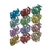
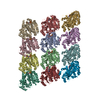
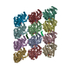
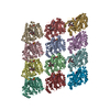
- PDB-6dpu: Undecorated GMPCPP microtubule -

+
Open data


ID or keywords:

Loading...

-
Basic information

Entry
Database: PDB / ID: 6dpu
TitleUndecorated GMPCPP microtubule
Components
  • Tubulin alpha-1B chain
  • Tubulin beta chain
KeywordsCELL CYCLE / microtubule / cytoskeleton / GMPCPP / seam
Function / homology
Function and homology information


Microtubule-dependent trafficking of connexons from Golgi to the plasma membrane / Hedgehog 'off' state / Cilium Assembly / Intraflagellar transport / COPI-dependent Golgi-to-ER retrograde traffic / Carboxyterminal post-translational modifications of tubulin / RHOH GTPase cycle / Sealing of the nuclear envelope (NE) by ESCRT-III / Kinesins / PKR-mediated signaling ...Microtubule-dependent trafficking of connexons from Golgi to the plasma membrane / Hedgehog 'off' state / Cilium Assembly / Intraflagellar transport / COPI-dependent Golgi-to-ER retrograde traffic / Carboxyterminal post-translational modifications of tubulin / RHOH GTPase cycle / Sealing of the nuclear envelope (NE) by ESCRT-III / Kinesins / PKR-mediated signaling / The role of GTSE1 in G2/M progression after G2 checkpoint / Aggrephagy / Resolution of Sister Chromatid Cohesion / Mitotic Prometaphase / EML4 and NUDC in mitotic spindle formation / Separation of Sister Chromatids / RHO GTPases activate IQGAPs / RHO GTPases Activate Formins / HSP90 chaperone cycle for steroid hormone receptors (SHR) in the presence of ligand / MHC class II antigen presentation / Recruitment of NuMA to mitotic centrosomes / COPI-mediated anterograde transport / structural constituent of cytoskeleton / microtubule cytoskeleton organization / microtubule cytoskeleton / mitotic cell cycle / Hydrolases; Acting on acid anhydrides; Acting on GTP to facilitate cellular and subcellular movement / microtubule / GTPase activity / GTP binding / metal ion binding / cytoplasm
Similarity search - Function
Helix hairpin bin / Tubulin/FtsZ, C-terminal domain / Tubulin/FtsZ, GTPase domain / 60s Ribosomal Protein L30; Chain: A; / Alpha tubulin / Tubulin-beta mRNA autoregulation signal. / Beta tubulin, autoregulation binding site / Beta tubulin / Tubulin / Tubulin, C-terminal ...Helix hairpin bin / Tubulin/FtsZ, C-terminal domain / Tubulin/FtsZ, GTPase domain / 60s Ribosomal Protein L30; Chain: A; / Alpha tubulin / Tubulin-beta mRNA autoregulation signal. / Beta tubulin, autoregulation binding site / Beta tubulin / Tubulin / Tubulin, C-terminal / Tubulin C-terminal domain / Tubulin, conserved site / Tubulin subunits alpha, beta, and gamma signature. / Tubulin/FtsZ family, C-terminal domain / Tubulin/FtsZ-like, C-terminal domain / Tubulin/FtsZ, C-terminal / Tubulin/FtsZ, 2-layer sandwich domain / Tubulin/FtsZ family, GTPase domain / Tubulin/FtsZ family, GTPase domain / Tubulin/FtsZ, GTPase domain / Tubulin/FtsZ, GTPase domain superfamily / Helix Hairpins / Rossmann fold / 2-Layer Sandwich / Orthogonal Bundle / 3-Layer(aba) Sandwich / Mainly Alpha / Alpha Beta
Similarity search - Domain/homology
PHOSPHOMETHYLPHOSPHONIC ACID GUANYLATE ESTER / GUANOSINE-5'-TRIPHOSPHATE / Tubulin beta chain / Tubulin alpha-1B chain
Similarity search - Component
Biological speciesSus scrofa (pig)
MethodELECTRON MICROSCOPY / helical reconstruction / cryo EM / Resolution: 3.1 Å
AuthorsZhang, R. / Nogales, E.
Funding support United States, 3items
OrganizationGrant numberCountry
National Institutes of Health/National Institute of General Medical Sciences (NIH/NIGMS)GM051487 United States
National Science Foundation (NSF, United States)1106400 United States
Howard Hughes Medical Institute (HHMI) United States
CitationJournal: Proc Natl Acad Sci U S A / Year: 2018
Title: Separating the effects of nucleotide and EB binding on microtubule structure.
Authors: Rui Zhang / Benjamin LaFrance / Eva Nogales /
Abstract: Microtubules (MTs) are polymers assembled from αβ-tubulin heterodimers that display the hallmark behavior of dynamic instability. MT dynamics are driven by GTP hydrolysis within the MT lattice, and ...Microtubules (MTs) are polymers assembled from αβ-tubulin heterodimers that display the hallmark behavior of dynamic instability. MT dynamics are driven by GTP hydrolysis within the MT lattice, and are highly regulated by a number of MT-associated proteins (MAPs). How MAPs affect MTs is still not fully understood, partly due to a lack of high-resolution structural data on undecorated MTs, which need to serve as a baseline for further comparisons. Here we report three structures of MTs in different nucleotide states (GMPCPP, GDP, and GTPγS) at near-atomic resolution and in the absence of any binding proteins. These structures allowed us to differentiate the effects of nucleotide state versus MAP binding on MT structure. Kinesin binding has a small effect on the extended, GMPCPP-bound lattice, but hardly affects the compacted GDP-MT lattice, while binding of end-binding (EB) proteins can induce lattice compaction (together with lattice twist) in MTs that were initially in an extended and more stable state. We propose a MT lattice-centric model in which the MT lattice serves as a platform that integrates internal tubulin signals, such as nucleotide state, with outside signals, such as binding of MAPs or mechanical forces, resulting in global lattice rearrangements that in turn affect the affinity of other MT partners and result in the exquisite regulation of MT dynamics.
History
DepositionJun 9, 2018Deposition site: RCSB / Processing site: RCSB
Revision 1.0Jul 4, 2018Provider: repository / Type: Initial release
Revision 1.1Jul 18, 2018Group: Data collection / Database references / Category: citation
Item: _citation.journal_volume / _citation.page_first / _citation.page_last
Revision 1.2Nov 20, 2019Group: Author supporting evidence / Category: pdbx_audit_support / Item: _pdbx_audit_support.funding_organization
Revision 1.3Mar 13, 2024Group: Data collection / Database references / Refinement description
Category: chem_comp_atom / chem_comp_bond ...chem_comp_atom / chem_comp_bond / database_2 / struct_ncs_dom_lim
Item: _database_2.pdbx_DOI / _database_2.pdbx_database_accession ..._database_2.pdbx_DOI / _database_2.pdbx_database_accession / _struct_ncs_dom_lim.beg_auth_comp_id / _struct_ncs_dom_lim.beg_label_asym_id / _struct_ncs_dom_lim.beg_label_comp_id / _struct_ncs_dom_lim.beg_label_seq_id / _struct_ncs_dom_lim.end_auth_comp_id / _struct_ncs_dom_lim.end_label_asym_id / _struct_ncs_dom_lim.end_label_comp_id / _struct_ncs_dom_lim.end_label_seq_id

-
Structure visualization

Movie
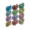
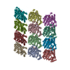
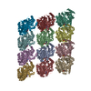
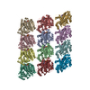
  • Deposited structure unit
  • Imaged by Jmol
  • Download
  • Simplified surface model + fitted atomic model
  • EMDB-7973
  • Imaged by Jmol
  • Download
  • Superimposition on EM map
  • EMDB-7973
  • Imaged by UCSF Chimera
  • Download
Movie viewer
Structure viewerMolecule:
MolmilJmol/JSmol

Downloads & links

-
Assembly

Deposited unit
A: Tubulin alpha-1B chain
B: Tubulin beta chain
C: Tubulin alpha-1B chain
D: Tubulin beta chain
E: Tubulin alpha-1B chain
F: Tubulin beta chain
G: Tubulin beta chain
H: Tubulin beta chain
I: Tubulin beta chain
J: Tubulin alpha-1B chain
K: Tubulin alpha-1B chain
L: Tubulin alpha-1B chain
hetero molecules


Theoretical massNumber of molelcules
Total (without water)607,23136
Polymers600,67312
Non-polymers6,55824
Water00
1


  • Idetical with deposited unit
  • defined by author
  • Evidence: microscopy
TypeNameSymmetry operationNumber
identity operation1_5551
Buried area50380 Å2
ΔGint-322 kcal/mol
Surface area168010 Å2
Noncrystallographic symmetry (NCS)NCS domain:
IDEns-IDDetails
11A
21C
12A
22E
13A
23J
14A
24K
15A
25L
16B
26D
17B
27F
18B
28G
19B
29H
110B
210I
111C
211E
112C
212J
113C
213K
114C
214L
115D
215F
116D
216G
117D
217H
118D
218I
119E
219J
120E
220K
121E
221L
122F
222G
123F
223H
124F
224I
125G
225H
126G
226I
127H
227I
128J
228K
129J
229L
130K
230L

NCS domain segments:

Component-ID: _ / Beg auth comp-ID: MET / Beg label comp-ID: MET / Refine code: _

Dom-IDEns-IDEnd auth comp-IDEnd label comp-IDAuth asym-IDLabel asym-IDAuth seq-IDLabel seq-ID
11SERSERAA1 - 4391 - 439
21SERSERCC1 - 4391 - 439
12SERSERAA1 - 4391 - 439
22SERSEREE1 - 4391 - 439
13SERSERAA1 - 4391 - 439
23SERSERJJ1 - 4391 - 439
14SERSERAA1 - 4391 - 439
24SERSERKK1 - 4391 - 439
15SERSERAA1 - 4391 - 439
25SERSERLL1 - 4391 - 439
16ASPASPBB1 - 4271 - 417
26ASPASPDD1 - 4271 - 417
17ASPASPBB1 - 4271 - 417
27ASPASPFF1 - 4271 - 417
18ASPASPBB1 - 4271 - 417
28ASPASPGG1 - 4271 - 417
19ASPASPBB1 - 4271 - 417
29ASPASPHH1 - 4271 - 417
110ASPASPBB1 - 4271 - 417
210ASPASPII1 - 4271 - 417
111SERSERCC1 - 4391 - 439
211SERSEREE1 - 4391 - 439
112SERSERCC1 - 4391 - 439
212SERSERJJ1 - 4391 - 439
113SERSERCC1 - 4391 - 439
213SERSERKK1 - 4391 - 439
114SERSERCC1 - 4391 - 439
214SERSERLL1 - 4391 - 439
115ASPASPDD1 - 4271 - 417
215ASPASPFF1 - 4271 - 417
116ASPASPDD1 - 4271 - 417
216ASPASPGG1 - 4271 - 417
117ASPASPDD1 - 4271 - 417
217ASPASPHH1 - 4271 - 417
118ASPASPDD1 - 4271 - 417
218ASPASPII1 - 4271 - 417
119SERSEREE1 - 4391 - 439
219SERSERJJ1 - 4391 - 439
120SERSEREE1 - 4391 - 439
220SERSERKK1 - 4391 - 439
121SERSEREE1 - 4391 - 439
221SERSERLL1 - 4391 - 439
122ASPASPFF1 - 4271 - 417
222ASPASPGG1 - 4271 - 417
123ASPASPFF1 - 4271 - 417
223ASPASPHH1 - 4271 - 417
124ASPASPFF1 - 4271 - 417
224ASPASPII1 - 4271 - 417
125ASPASPGG1 - 4271 - 417
225ASPASPHH1 - 4271 - 417
126ASPASPGG1 - 4271 - 417
226ASPASPII1 - 4271 - 417
127ASPASPHH1 - 4271 - 417
227ASPASPII1 - 4271 - 417
128SERSERJJ1 - 4391 - 439
228SERSERKK1 - 4391 - 439
129SERSERJJ1 - 4391 - 439
229SERSERLL1 - 4391 - 439
130SERSERKK1 - 4391 - 439
230SERSERLL1 - 4391 - 439

NCS ensembles :
ID
1
2
3
4
5
6
7
8
9
10
11
12
13
14
15
16
17
18
19
20
21
22
23
24
25
26
27
28
29
30

-
Components

#1: Protein
Tubulin alpha-1B chain / Alpha-tubulin ubiquitous / Tubulin K-alpha-1 / Tubulin alpha-ubiquitous chain


Mass: 50204.445 Da / Num. of mol.: 6 / Source method: isolated from a natural source / Source: (natural) Sus scrofa (pig) / References: UniProt: Q2XVP4
#2: Protein
Tubulin beta chain / Beta-tubulin


Mass: 49907.770 Da / Num. of mol.: 6 / Source method: isolated from a natural source / Source: (natural) Sus scrofa (pig) / References: UniProt: P02554
#3: Chemical
ChemComp-GTP / GUANOSINE-5'-TRIPHOSPHATE


Mass: 523.180 Da / Num. of mol.: 6 / Source method: obtained synthetically / Formula: C10H16N5O14P3 / Comment: GTP, energy-carrying molecule*YM
#4: Chemical
ChemComp-MG / MAGNESIUM ION


Mass: 24.305 Da / Num. of mol.: 12 / Source method: obtained synthetically / Formula: Mg
#5: Chemical
ChemComp-G2P / PHOSPHOMETHYLPHOSPHONIC ACID GUANYLATE ESTER


Mass: 521.208 Da / Num. of mol.: 6 / Source method: obtained synthetically / Formula: C11H18N5O13P3 / Comment: GMP-CPP, energy-carrying molecule analogue*YM

-
Experimental details

-
Experiment

ExperimentMethod: ELECTRON MICROSCOPY
EM experimentAggregation state: HELICAL ARRAY / 3D reconstruction method: helical reconstruction

-
Sample preparation

ComponentName: microtubule stabilized by GMPCPP / Type: COMPLEX / Entity ID: #1-#2 / Source: NATURAL
Molecular weightExperimental value: NO
Source (natural)Organism: Sus scrofa (pig)
Buffer solutionpH: 6.8
Details: BRB80 buffer (80 mM PIPES, pH 6.8, 1 mM EGTA, 1 mM MgCl2) supplemented with 1 mM GMPCPP, 1 mM DTT, and 0.05% Nonident P-40
SpecimenConc.: 3 mg/ml / Embedding applied: NO / Shadowing applied: NO / Staining applied: NO / Vitrification applied: YES
Details: Microtubules were polymerized at 37 degrees for 30 minutes.
Specimen supportDetails: Glow discharge was performed with Pelco EasiGlow device.
Grid material: COPPER / Grid mesh size: 400 divisions/in. / Grid type: C-flat-1.2/1.3 4C
VitrificationInstrument: FEI VITROBOT MARK IV / Cryogen name: ETHANE / Humidity: 95 % / Chamber temperature: 298 K / Details: Blot for 4 seconds before plunging.

-
Electron microscopy imaging

Experimental equipment
Model: Titan Krios / Image courtesy: FEI Company
MicroscopyModel: FEI TITAN KRIOS
Electron gunElectron source: FIELD EMISSION GUN / Accelerating voltage: 300 kV / Illumination mode: FLOOD BEAM
Electron lensMode: BRIGHT FIELD / Nominal magnification: 105000 X / Nominal defocus max: 2500 nm / Nominal defocus min: 1000 nm / Calibrated defocus min: 800 nm / Calibrated defocus max: 2700 nm / Cs: 0.01 mm / Alignment procedure: COMA FREE
Specimen holderCryogen: NITROGEN / Specimen holder model: FEI TITAN KRIOS AUTOGRID HOLDER
Image recordingAverage exposure time: 8 sec. / Electron dose: 1.33 e/Å2 / Detector mode: COUNTING / Film or detector model: GATAN K2 QUANTUM (4k x 4k) / Num. of grids imaged: 1 / Num. of real images: 1176
EM imaging opticsEnergyfilter name: GIF Bioquantum / Energyfilter slit width: 20 eV
Spherical aberration corrector: Microscope has a Cs corrector.
Image scansWidth: 3838 / Height: 3710 / Movie frames/image: 40 / Used frames/image: 1-40

-
Processing

SoftwareName: REFMAC / Version: 5.8.0091 / Classification: refinement
EM software
IDNameCategory
1Appionparticle selection
2EPUimage acquisition
4GctfCTF correction
7UCSF Chimeramodel fitting
9REFMACmodel refinement
10EMANinitial Euler assignment
11FREALIGNfinal Euler assignment
13FREALIGN3D reconstruction
CTF correctionType: PHASE FLIPPING AND AMPLITUDE CORRECTION
Helical symmertyAngular rotation/subunit: -25.75 ° / Axial rise/subunit: 8.99 Å / Axial symmetry: C1
3D reconstructionResolution: 3.1 Å / Resolution method: FSC 0.143 CUT-OFF / Num. of particles: 110127 / Algorithm: FOURIER SPACE / Num. of class averages: 1 / Symmetry type: HELICAL
Atomic model buildingB value: 150 / Protocol: RIGID BODY FIT / Space: REAL / Target criteria: Correlation coefficient
RefinementResolution: 3.1→214.82 Å / Cor.coef. Fo:Fc: 0.768 / SU B: 32.588 / SU ML: 0.54 / ESU R: 0.978
Stereochemistry target values: MAXIMUM LIKELIHOOD WITH PHASES
Details: HYDROGENS HAVE BEEN ADDED IN THE RIDING POSITIONS
RfactorNum. reflection% reflection
Rwork0.41337 --
obs0.41337 232359 100 %
Solvent computationSolvent model: PARAMETERS FOR MASK CACLULATION
Displacement parametersBiso mean: 29.477 Å2
Baniso -1Baniso -2Baniso -3
1--3.04 Å2-0.23 Å2-0.3 Å2
2--8.45 Å2-0.38 Å2
3----5.41 Å2
Refinement stepCycle: 1 / Total: 40650
Refine LS restraints
Refine-IDTypeDev idealDev ideal targetNumber
ELECTRON MICROSCOPYr_bond_refined_d0.0080.01941574
ELECTRON MICROSCOPYr_bond_other_d0.0020.0238316
ELECTRON MICROSCOPYr_angle_refined_deg1.4451.95956490
ELECTRON MICROSCOPYr_angle_other_deg0.972388134
ELECTRON MICROSCOPYr_dihedral_angle_1_deg12.2044.8365813
ELECTRON MICROSCOPYr_dihedral_angle_2_deg35.06824.2131956
ELECTRON MICROSCOPYr_dihedral_angle_3_deg16.033156693
ELECTRON MICROSCOPYr_dihedral_angle_4_deg11.90115251
ELECTRON MICROSCOPYr_chiral_restr0.0820.26174
ELECTRON MICROSCOPYr_gen_planes_refined0.0050.02147472
ELECTRON MICROSCOPYr_gen_planes_other0.0020.029792
ELECTRON MICROSCOPYr_nbd_refined
ELECTRON MICROSCOPYr_nbd_other
ELECTRON MICROSCOPYr_nbtor_refined
ELECTRON MICROSCOPYr_nbtor_other
ELECTRON MICROSCOPYr_xyhbond_nbd_refined
ELECTRON MICROSCOPYr_xyhbond_nbd_other
ELECTRON MICROSCOPYr_metal_ion_refined
ELECTRON MICROSCOPYr_metal_ion_other
ELECTRON MICROSCOPYr_symmetry_vdw_refined
ELECTRON MICROSCOPYr_symmetry_vdw_other
ELECTRON MICROSCOPYr_symmetry_hbond_refined
ELECTRON MICROSCOPYr_symmetry_hbond_other
ELECTRON MICROSCOPYr_symmetry_metal_ion_refined
ELECTRON MICROSCOPYr_symmetry_metal_ion_other
ELECTRON MICROSCOPYr_mcbond_it0.9472.92820550
ELECTRON MICROSCOPYr_mcbond_other0.9472.92820549
ELECTRON MICROSCOPYr_mcangle_it1.7714.38825656
ELECTRON MICROSCOPYr_mcangle_other1.7714.38825657
ELECTRON MICROSCOPYr_scbond_it0.6953.04121024
ELECTRON MICROSCOPYr_scbond_other0.6953.04221012
ELECTRON MICROSCOPYr_scangle_it
ELECTRON MICROSCOPYr_scangle_other1.364.49530815
ELECTRON MICROSCOPYr_long_range_B_refined4.30226.0195315
ELECTRON MICROSCOPYr_long_range_B_other4.30226.01595297
ELECTRON MICROSCOPYr_rigid_bond_restr
ELECTRON MICROSCOPYr_sphericity_free
ELECTRON MICROSCOPYr_sphericity_bonded
Refine LS restraints NCS

Refine-ID: ELECTRON MICROSCOPY / Type: interatomic distance / Rms dev position: 0 Å / Weight position: 0.05

Ens-IDDom-IDAuth asym-IDNumber
11A52726
12C52726
21A52728
22E52728
31A52718
32J52718
41A52720
42K52720
51A52718
52L52718
61B52638
62D52638
71B52638
72F52638
81B52636
82G52636
91B52634
92H52634
101B52632
102I52632
111C52728
112E52728
121C52718
122J52718
131C52724
132K52724
141C52722
142L52722
151D52644
152F52644
161D52642
162G52642
171D52640
172H52640
181D52640
182I52640
191E52724
192J52724
201E52722
202K52722
211E52720
212L52720
221F52648
222G52648
231F52642
232H52642
241F52640
242I52640
251G52642
252H52642
261G52640
262I52640
271H52640
272I52640
281J52720
282K52720
291J52722
292L52722
301K52724
302L52724
LS refinement shellResolution: 3.1→3.18 Å / Total num. of bins used: 20
RfactorNum. reflection% reflection
Rwork0.736 17224 -
Rfree-0 -
obs--100 %

+
About Yorodumi

-
News

-
Feb 9, 2022. New format data for meta-information of EMDB entries

New format data for meta-information of EMDB entries

  • Version 3 of the EMDB header file is now the official format.
  • The previous official version 1.9 will be removed from the archive.

Related info.:EMDB header

External links:wwPDB to switch to version 3 of the EMDB data model

-
Aug 12, 2020. Covid-19 info

Covid-19 info

URL: https://pdbj.org/emnavi/covid19.php

New page: Covid-19 featured information page in EM Navigator.

Related info.:Covid-19 info / Mar 5, 2020. Novel coronavirus structure data

+
Mar 5, 2020. Novel coronavirus structure data

Novel coronavirus structure data

Related info.:Yorodumi Speices / Aug 12, 2020. Covid-19 info

External links:COVID-19 featured content - PDBj / Molecule of the Month (242):Coronavirus Proteases

+
Jan 31, 2019. EMDB accession codes are about to change! (news from PDBe EMDB page)

EMDB accession codes are about to change! (news from PDBe EMDB page)

  • The allocation of 4 digits for EMDB accession codes will soon come to an end. Whilst these codes will remain in use, new EMDB accession codes will include an additional digit and will expand incrementally as the available range of codes is exhausted. The current 4-digit format prefixed with “EMD-” (i.e. EMD-XXXX) will advance to a 5-digit format (i.e. EMD-XXXXX), and so on. It is currently estimated that the 4-digit codes will be depleted around Spring 2019, at which point the 5-digit format will come into force.
  • The EM Navigator/Yorodumi systems omit the EMD- prefix.

Related info.:Q: What is EMD? / ID/Accession-code notation in Yorodumi/EM Navigator

External links:EMDB Accession Codes are Changing Soon! / Contact to PDBj

+
Jul 12, 2017. Major update of PDB

Major update of PDB

  • wwPDB released updated PDB data conforming to the new PDBx/mmCIF dictionary.
  • This is a major update changing the version number from 4 to 5, and with Remediation, in which all the entries are updated.
  • In this update, many items about electron microscopy experimental information are reorganized (e.g. em_software).
  • Now, EM Navigator and Yorodumi are based on the updated data.

External links:wwPDB Remediation / Enriched Model Files Conforming to OneDep Data Standards Now Available in the PDB FTP Archive

-
Yorodumi

Thousand views of thousand structures

  • Yorodumi is a browser for structure data from EMDB, PDB, SASBDB, etc.
  • This page is also the successor to EM Navigator detail page, and also detail information page/front-end page for Omokage search.
  • The word "yorodu" (or yorozu) is an old Japanese word meaning "ten thousand". "mi" (miru) is to see.

Related info.:EMDB / PDB / SASBDB / Comparison of 3 databanks / Yorodumi Search / Aug 31, 2016. New EM Navigator & Yorodumi / Yorodumi Papers / Jmol/JSmol / Function and homology information / Changes in new EM Navigator and Yorodumi

Read more