 Movie
Movie Controller


+ Open data

- Basic information
| Entry | Database: PDB / ID: 6.0E+88 | ||||||||||||
|---|---|---|---|---|---|---|---|---|---|---|---|---|---|
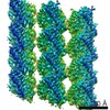
| Title | Cryo-EM structure of C. elegans GDP-microtubule | ||||||||||||
 Components Components |
| ||||||||||||
 Keywords Keywords | STRUCTURAL PROTEIN / Cytoskeletal protein | ||||||||||||
| Function / homology |  Function and homology information Function and homology informationCOPI-mediated anterograde transport / COPI-independent Golgi-to-ER retrograde traffic / Intraflagellar transport / Neutrophil degranulation / meiotic spindle organization / embryo development ending in birth or egg hatching / centrosome localization / tubulin complex / establishment of mitotic spindle orientation / regulation of cytokinesis ...COPI-mediated anterograde transport / COPI-independent Golgi-to-ER retrograde traffic / Intraflagellar transport / Neutrophil degranulation / meiotic spindle organization / embryo development ending in birth or egg hatching / centrosome localization / tubulin complex / establishment of mitotic spindle orientation / regulation of cytokinesis / spindle microtubule / microtubule cytoskeleton organization / structural constituent of cytoskeleton / intracellular protein localization / mitotic cell cycle / microtubule / Hydrolases; Acting on acid anhydrides; Acting on GTP to facilitate cellular and subcellular movement / hydrolase activity / GTPase activity / GTP binding / metal ion binding / cytoplasm Similarity search - Function | ||||||||||||
| Biological species |  | ||||||||||||
| Method | ELECTRON MICROSCOPY / single particle reconstruction / cryo EM / Resolution: 4.8 Å | ||||||||||||
 Authors Authors | Chaaban, S. / Jariwala, S. / Chieh-Ting, H. / Redemann, S. / Kollman, J. / Muller-Reichert, T. / Sept, D. / Bui, K.H. / Brouhard, G.J. | ||||||||||||
| Funding support |  Canada, 3items Canada, 3items
| ||||||||||||
 Citation Citation | Journal: Dev Cell / Year: 2018 Title: The Structure and Dynamics of C. elegans Tubulin Reveals the Mechanistic Basis of Microtubule Growth. Authors: Sami Chaaban / Shashank Jariwala / Chieh-Ting Hsu / Stefanie Redemann / Justin M Kollman / Thomas Müller-Reichert / David Sept / Khanh Huy Bui / Gary J Brouhard /   Abstract: The dynamic instability of microtubules is a conserved and fundamental mechanism in eukaryotes. Yet microtubules from different species diverge in their growth rates, lattice structures, and ...The dynamic instability of microtubules is a conserved and fundamental mechanism in eukaryotes. Yet microtubules from different species diverge in their growth rates, lattice structures, and responses to GTP hydrolysis. Therefore, we do not know what limits microtubule growth, what determines microtubule structure, or whether the mechanisms of dynamic instability are universal. Here, we studied microtubules from the nematode C. elegans, which have strikingly fast growth rates and non-canonical lattices in vivo. Using a reconstitution approach, we discovered that C. elegans microtubules combine intrinsically fast growth with very frequent catastrophes. We solved the structure of C. elegans microtubules to 4.8 Å and discovered sequence divergence in the lateral contact loops, one of which is ordered in C. elegans but unresolved in other species. We provide direct evidence that C. elegans tubulin has a higher free energy in solution and propose a model wherein the ordering of lateral contact loops activates tubulin for growth. | ||||||||||||
| History |
|
- Structure visualization
| Movie |
Movie viewer |
|---|---|
| Structure viewer | Molecule: MolmilJmol/JSmol |
- Downloads & links
-Download
| PDBx/mmCIF format | 6e88.cif.gz | 876.2 KB | Display | PDBx/mmCIF format |
|---|---|---|---|---|
| PDB format | pdb6e88.ent.gz | 733.5 KB | Display | PDB format |
| PDBx/mmJSON format | 6e88.json.gz | Tree view | PDBx/mmJSON format | |
| Others |  Other downloads Other downloads |
-Validation report
| Arichive directory | https://data.pdbj.org/pub/pdb/validation_reports/e8/6e88ftp://data.pdbj.org/pub/pdb/validation_reports/e8/6e88 | HTTPS FTP |
|---|
-Related structure data
| Related structure data |  9004MC M: map data used to model this data C: citing same article ( |
|---|---|
| Similar structure data |

-Links
 PDBj
PDBj


- Assembly
| Deposited unit | 
|
|---|---|
| 1 |
|
-Components
| #1: Protein | Mass: 48467.609 Da / Num. of mol.: 6 / Source method: isolated from a natural source / Source: (natural) #2: Protein | Mass: 47843.988 Da / Num. of mol.: 6 / Source method: isolated from a natural source / Source: (natural) #3: Chemical | ChemComp-GTP /   Mass: 523.180 Da / Num. of mol.: 6 / Source method: obtained synthetically / Formula: C10H16N5O14P3 / Comment: GTP, energy-carrying molecule*YM Mass: 523.180 Da / Num. of mol.: 6 / Source method: obtained synthetically / Formula: C10H16N5O14P3 / Comment: GTP, energy-carrying molecule*YM#4: Chemical | ChemComp-GDP /   Type: RNA linking / Mass: 443.201 Da / Num. of mol.: 6 / Source method: obtained synthetically / Formula: C10H15N5O11P2 / Comment: GDP, energy-carrying molecule*YM Type: RNA linking / Mass: 443.201 Da / Num. of mol.: 6 / Source method: obtained synthetically / Formula: C10H15N5O11P2 / Comment: GDP, energy-carrying molecule*YM |
|---|
-Experimental details
-Experiment
| Experiment | Method: ELECTRON MICROSCOPY |
|---|---|
| EM experiment | Aggregation state: FILAMENT / 3D reconstruction method: single particle reconstruction |
- Sample preparation
| Component | Name: microtubule / Type: ORGANELLE OR CELLULAR COMPONENT Details: C. elegans microtubules polymerized in the presence of GTP Entity ID: #1-#2 / Source: NATURAL |
|---|---|
| Molecular weight | Value: 165 kDa/nm / Experimental value: NO |
| Source (natural) | Organism: |
| Buffer solution | pH: 6.9 |
| Specimen | Embedding applied: NO / Shadowing applied: NO / Staining applied: NO / Vitrification applied: YES |
| Specimen support | Details: unspecified |
| Vitrification | Cryogen name: ETHANE |
- Electron microscopy imaging
| Experimental equipment |  Model: Titan Krios / Image courtesy: FEI Company |
|---|---|
| Microscopy | Model: FEI TITAN KRIOS |
| Electron gun | Electron source:  FIELD EMISSION GUN / Accelerating voltage: 300 kV / Illumination mode: SPOT SCAN FIELD EMISSION GUN / Accelerating voltage: 300 kV / Illumination mode: SPOT SCAN |
| Electron lens | Mode: BRIGHT FIELD |
| Image recording | Electron dose: 29.33 e/Å2 / Film or detector model: FEI FALCON II (4k x 4k) |
- Processing
| CTF correction | Type: PHASE FLIPPING AND AMPLITUDE CORRECTION |
|---|---|
| 3D reconstruction | Resolution: 4.8 Å / Resolution method: FSC 0.143 CUT-OFF / Num. of particles: 16574 / Symmetry type: POINT |
| Atomic model building | Protocol: FLEXIBLE FIT |
