 Movie
Movie Controller

[English] 日本語
 Yorodumi
- PDB-6d5f: Cryo-EM reconstruction of membrane-enveloped filamentous virus SF... -
+ Open data

- Basic information
| Entry | Database: PDB / ID: 6d5f | ||||||
|---|---|---|---|---|---|---|---|
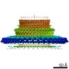
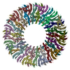
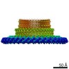
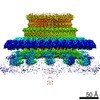
| Title | Cryo-EM reconstruction of membrane-enveloped filamentous virus SFV1 (Sulfolobus filamentous virus 1) | ||||||
 Components Components |
| ||||||
 Keywords Keywords | VIRUS / filamentous virus / helical symmetry / membrane enveloped virus | ||||||
| Function / homology | DNA / DNA (> 10) / DNA (> 100) Function and homology information Function and homology information | ||||||
| Biological species |   Sulfolobus filamentous virus 1 Sulfolobus filamentous virus 1 | ||||||
| Method | ELECTRON MICROSCOPY / helical reconstruction / cryo EM / Resolution: 3.7 Å | ||||||
 Authors Authors | Wang, F. / Osinski, T. / Liu, Y. / Krupovic, M. / Prangishvili, D. / Egelman, E.H. | ||||||
| Funding support |  United States, 1items United States, 1items
| ||||||
 Citation Citation | Journal: Nat Commun / Year: 2018 Title: Structural conservation in a membrane-enveloped filamentous virus infecting a hyperthermophilic acidophile. Authors: Ying Liu / Tomasz Osinski / Fengbin Wang / Mart Krupovic / Stefan Schouten / Peter Kasson / David Prangishvili / Edward H Egelman /   Abstract: Different forms of viruses that infect archaea inhabiting extreme environments continue to be discovered at a surprising rate, suggesting that the current sampling of these viruses is sparse. We ...Different forms of viruses that infect archaea inhabiting extreme environments continue to be discovered at a surprising rate, suggesting that the current sampling of these viruses is sparse. We describe here Sulfolobus filamentous virus 1 (SFV1), a membrane-enveloped virus infecting Sulfolobus shibatae. The virus encodes two major coat proteins which display no apparent sequence similarity with each other or with any other proteins in databases. We have used cryo-electron microscopy at 3.7 Å resolution to show that these two proteins form a nearly symmetrical heterodimer, which wraps around A-form DNA, similar to what has been shown for SIRV2 and AFV1, two other archaeal filamentous viruses. The thin (∼ 20 Å) membrane of SFV1 is mainly archaeol, a lipid species that accounts for only 1% of the host lipids. Our results show how relatively conserved structural features can be maintained across evolution by both proteins and lipids that have diverged considerably. | ||||||
| History |
|
- Structure visualization
| Movie |
Movie viewer |
|---|---|
| Structure viewer | Molecule: MolmilJmol/JSmol |
- Downloads & links
-Download
| PDBx/mmCIF format | 6d5f.cif.gz | 1.6 MB | Display | PDBx/mmCIF format |
|---|---|---|---|---|
| PDB format | pdb6d5f.ent.gz | 1.4 MB | Display | PDB format |
| PDBx/mmJSON format | 6d5f.json.gz | Tree view | PDBx/mmJSON format | |
| Others |  Other downloads Other downloads |
-Validation report
| Arichive directory | https://data.pdbj.org/pub/pdb/validation_reports/d5/6d5fftp://data.pdbj.org/pub/pdb/validation_reports/d5/6d5f | HTTPS FTP |
|---|
-Related structure data
| Related structure data |  7797MC M: map data used to model this data C: citing same article ( |
|---|---|
| Similar structure data |


-Links
 PDBj
PDBj


- Assembly
| Deposited unit | 
|
|---|---|
| 1 |
|
-Components
| #1: Protein | Mass: 22020.135 Da / Num. of mol.: 26 / Source method: isolated from a natural source / Source: (natural) Sulfolobus filamentous virus 1#2: Protein | Mass: 15792.202 Da / Num. of mol.: 26 / Source method: isolated from a natural source / Source: (natural) Sulfolobus filamentous virus 1#3: DNA chain | Mass: 103678.133 Da / Num. of mol.: 2 / Source method: isolated from a natural source / Source: (natural) Sulfolobus filamentous virus 1Has protein modification | Y | |
|---|
-Experimental details
-Experiment
| Experiment | Method: ELECTRON MICROSCOPY |
|---|---|
| EM experiment | Aggregation state: FILAMENT / 3D reconstruction method: helical reconstruction |
- Sample preparation
| Component | Name: Sulfolobus filamentous virus 1 / Type: VIRUS / Entity ID: all / Source: NATURAL |
|---|---|
| Source (natural) | Organism: Sulfolobus filamentous virus 1 |
| Details of virus | Empty: NO / Enveloped: YES / Isolate: STRAIN / Type: VIRION |
| Buffer solution | pH: 6 / Details: 20 mM Tris-acetate, 250 mM sodium chloride, pH 6.0 |
| Specimen | Embedding applied: NO / Shadowing applied: NO / Staining applied: NO / Vitrification applied: YES |
| Vitrification | Cryogen name: ETHANE / Humidity: 90 % |
- Electron microscopy imaging
| Experimental equipment |  Model: Titan Krios / Image courtesy: FEI Company |
|---|---|
| Microscopy | Model: FEI TITAN KRIOS |
| Electron gun | Electron source:  FIELD EMISSION GUN / Accelerating voltage: 300 kV / Illumination mode: FLOOD BEAM FIELD EMISSION GUN / Accelerating voltage: 300 kV / Illumination mode: FLOOD BEAM |
| Electron lens | Mode: BRIGHT FIELD |
| Image recording | Average exposure time: 2 sec. / Electron dose: 48 e/Å2 / Detector mode: INTEGRATING / Film or detector model: FEI FALCON III (4k x 4k) Details: Images were stored containing 24 fractions, where each fraction corresponded to a dose of ~2 electrons per Angstrom^2. |
- Processing
| Software | Name: PHENIX / Version: dev_2919: / Classification: refinement | ||||||||||||||||||||||||||||||
|---|---|---|---|---|---|---|---|---|---|---|---|---|---|---|---|---|---|---|---|---|---|---|---|---|---|---|---|---|---|---|---|
| EM software |
| ||||||||||||||||||||||||||||||
| CTF correction | Type: PHASE FLIPPING AND AMPLITUDE CORRECTION | ||||||||||||||||||||||||||||||
| Helical symmerty | Angular rotation/subunit: 21.02572 ° / Axial rise/subunit: 2.76412 Å / Axial symmetry: C1 | ||||||||||||||||||||||||||||||
| 3D reconstruction | Resolution: 3.7 Å / Resolution method: FSC 0.143 CUT-OFF / Num. of particles: 87803 / Symmetry type: HELICAL | ||||||||||||||||||||||||||||||
| Refinement | Highest resolution: 3.7 Å |
