 Movie
Movie Controller

[English] 日本語
 Yorodumi
- EMDB-7797: Cryo-EM reconstruction of membrane-enveloped filamentous virus SF... -
+ Open data

- Basic information
| Entry | Database: EMDB / ID: EMD-7797 | |||||||||
|---|---|---|---|---|---|---|---|---|---|---|
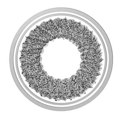
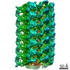
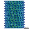
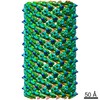
| Title | Cryo-EM reconstruction of membrane-enveloped filamentous virus SFV1 (Sulfolobus filamentous virus 1) | |||||||||
 Map data Map data | membrane-enveloped filamentous virus SFV1 | |||||||||
 Sample Sample |
| |||||||||
 Keywords Keywords | filamentous virus / helical symmetry / membrane enveloped virus / VIRUS | |||||||||
| Biological species |   Sulfolobus filamentous virus 1 Sulfolobus filamentous virus 1 | |||||||||
| Method | helical reconstruction / cryo EM / Resolution: 3.7 Å | |||||||||
 Authors Authors | Wang F / Osinski T | |||||||||
| Funding support |  United States, 1 items United States, 1 items
| |||||||||
 Citation Citation | Journal: Nat Commun / Year: 2018 Title: Structural conservation in a membrane-enveloped filamentous virus infecting a hyperthermophilic acidophile. Authors: Ying Liu / Tomasz Osinski / Fengbin Wang / Mart Krupovic / Stefan Schouten / Peter Kasson / David Prangishvili / Edward H Egelman /   Abstract: Different forms of viruses that infect archaea inhabiting extreme environments continue to be discovered at a surprising rate, suggesting that the current sampling of these viruses is sparse. We ...Different forms of viruses that infect archaea inhabiting extreme environments continue to be discovered at a surprising rate, suggesting that the current sampling of these viruses is sparse. We describe here Sulfolobus filamentous virus 1 (SFV1), a membrane-enveloped virus infecting Sulfolobus shibatae. The virus encodes two major coat proteins which display no apparent sequence similarity with each other or with any other proteins in databases. We have used cryo-electron microscopy at 3.7 Å resolution to show that these two proteins form a nearly symmetrical heterodimer, which wraps around A-form DNA, similar to what has been shown for SIRV2 and AFV1, two other archaeal filamentous viruses. The thin (∼ 20 Å) membrane of SFV1 is mainly archaeol, a lipid species that accounts for only 1% of the host lipids. Our results show how relatively conserved structural features can be maintained across evolution by both proteins and lipids that have diverged considerably. | |||||||||
| History |
|
- Structure visualization
| Movie |
 Movie viewer Movie viewer |
|---|---|
| Structure viewer | EM map: SurfViewMolmilJmol/JSmol |
| Supplemental images |
- Downloads & links
-EMDB archive
| Map data | emd_7797.map.gz | 195.3 MB | EMDB map data format | |
|---|---|---|---|---|
| Header (meta data) | emd-7797-v30.xmlemd-7797.xml | 12.9 KB 12.9 KB | Display Display | EMDB header |
| Images |  emd_7797.png emd_7797.png | 153.7 KB | ||
| Filedesc metadata | emd-7797.cif.gz | 5.3 KB | ||
| Archive directory |  http://ftp.pdbj.org/pub/emdb/structures/EMD-7797ftp://ftp.pdbj.org/pub/emdb/structures/EMD-7797 http://ftp.pdbj.org/pub/emdb/structures/EMD-7797ftp://ftp.pdbj.org/pub/emdb/structures/EMD-7797 | HTTPS FTP |
-Related structure data
| Related structure data |  6d5fMC M: atomic model generated by this map C: citing same article ( |
|---|---|
| Similar structure data |
-Links
| EMDB pages | EMDB (EBI/PDBe) / EMDataResource |
|---|
-Map
| File | Download / File: emd_7797.map.gz / Format: CCP4 / Size: 216 MB / Type: IMAGE STORED AS FLOATING POINT NUMBER (4 BYTES) | ||||||||||||||||||||||||||||||||||||||||||||||||||||||||||||||||||||
|---|---|---|---|---|---|---|---|---|---|---|---|---|---|---|---|---|---|---|---|---|---|---|---|---|---|---|---|---|---|---|---|---|---|---|---|---|---|---|---|---|---|---|---|---|---|---|---|---|---|---|---|---|---|---|---|---|---|---|---|---|---|---|---|---|---|---|---|---|---|
| Annotation | membrane-enveloped filamentous virus SFV1 | ||||||||||||||||||||||||||||||||||||||||||||||||||||||||||||||||||||
| Projections & slices | Image control
Images are generated by Spider. | ||||||||||||||||||||||||||||||||||||||||||||||||||||||||||||||||||||
| Voxel size | X=Y=Z: 1.09 Å | ||||||||||||||||||||||||||||||||||||||||||||||||||||||||||||||||||||
| Density |
| ||||||||||||||||||||||||||||||||||||||||||||||||||||||||||||||||||||
| Symmetry | Space group: 1 | ||||||||||||||||||||||||||||||||||||||||||||||||||||||||||||||||||||
| Details | EMDB XML:
CCP4 map header:
| ||||||||||||||||||||||||||||||||||||||||||||||||||||||||||||||||||||
 Z (Sec.)
 Y (Row.)
 X (Col.)


-Supplemental data
- Sample components
-Entire : Sulfolobus filamentous virus 1
| Entire | Name: Sulfolobus filamentous virus 1 |
|---|---|
| Components |
|
-Supramolecule #1: Sulfolobus filamentous virus 1
| Supramolecule | Name: Sulfolobus filamentous virus 1 / type: virus / ID: 1 / Parent: 0 / Macromolecule list: all / NCBI-ID: 2304198 / Sci species name: Sulfolobus filamentous virus 1 / Virus type: VIRION / Virus isolate: STRAIN / Virus enveloped: Yes / Virus empty: No |
|---|
-Macromolecule #1: Fimbrial protein
| Macromolecule | Name: Fimbrial protein / type: protein_or_peptide / ID: 1 / Number of copies: 26 / Enantiomer: LEVO |
|---|---|
| Source (natural) | Organism: Sulfolobus filamentous virus 1 |
| Molecular weight | Theoretical: 22.020135 KDa |
| Sequence | String: MARKRTSKND PLRMYLNYVR KLQTMGDAYD ESAKYRIANF ENGFKSLHMV ENEFKQYLAN VIDEAIKSGA SPQDLPYVNE IKLALMKIF TSWLKYSNEK LGANEIAINV AGTATMTLTE NLYGTRVSCE EAVSLINSIF AVWVGVEPFE AEEREGACLV T PRSPLPPV ...String: MARKRTSKND PLRMYLNYVR KLQTMGDAYD ESAKYRIANF ENGFKSLHMV ENEFKQYLAN VIDEAIKSGA SPQDLPYVNE IKLALMKIF TSWLKYSNEK LGANEIAINV AGTATMTLTE NLYGTRVSCE EAVSLINSIF AVWVGVEPFE AEEREGACLV T PRSPLPPV PISSPTGFSA PIQEVLQAKS PEEIIGVKGG A |
-Macromolecule #2: Fimbrial protein
| Macromolecule | Name: Fimbrial protein / type: protein_or_peptide / ID: 2 / Number of copies: 26 / Enantiomer: LEVO |
|---|---|
| Source (natural) | Organism: Sulfolobus filamentous virus 1 |
| Molecular weight | Theoretical: 15.792202 KDa |
| Sequence | String: MPSRRTGITT EDAITKYSVK AKTEQTAYKN ATKDMVVQAQ NIMNFYSVVN QALIPWLNAH GVGGNLRILY RQLANEYVKV LNTKQSGEV IKRLKIALRH KYWLRGLDEA MLDEFMDYID SLKSTTTNYI IFNMQSSK |
-Macromolecule #3: DNA (336-MER)
| Macromolecule | Name: DNA (336-MER) / type: dna / ID: 3 / Number of copies: 2 / Classification: DNA |
|---|---|
| Source (natural) | Organism: Sulfolobus filamentous virus 1 |
| Molecular weight | Theoretical: 103.678133 KDa |
| Sequence | String: (DT)(DA)(DT)(DA)(DT)(DA)(DT)(DA)(DT)(DA) (DT)(DA)(DT)(DA)(DT)(DA)(DT)(DA)(DT)(DA) (DT)(DA)(DT)(DA)(DT)(DA)(DT)(DA)(DT) (DA)(DT)(DA)(DT)(DA)(DT)(DA)(DT)(DA)(DT) (DA) (DT)(DA)(DT)(DA)(DT)(DA) ...String: (DT)(DA)(DT)(DA)(DT)(DA)(DT)(DA)(DT)(DA) (DT)(DA)(DT)(DA)(DT)(DA)(DT)(DA)(DT)(DA) (DT)(DA)(DT)(DA)(DT)(DA)(DT)(DA)(DT) (DA)(DT)(DA)(DT)(DA)(DT)(DA)(DT)(DA)(DT) (DA) (DT)(DA)(DT)(DA)(DT)(DA)(DT)(DA) (DT)(DA)(DT)(DA)(DT)(DA)(DT)(DA)(DT)(DA) (DT)(DA) (DT)(DA)(DT)(DA)(DT)(DA)(DT) (DA)(DT)(DA)(DT)(DA)(DT)(DA)(DT)(DA)(DT) (DA)(DT)(DA) (DT)(DA)(DT)(DA)(DT)(DA) (DT)(DA)(DT)(DA)(DT)(DA)(DT)(DA)(DT)(DA) (DT)(DA)(DT)(DA) (DT)(DA)(DT)(DA)(DT) (DA)(DT)(DA)(DT)(DA)(DT)(DA)(DT)(DA)(DT) (DA)(DT)(DA)(DT)(DA) (DT)(DA)(DT)(DA) (DT)(DA)(DT)(DA)(DT)(DA)(DT)(DA)(DT)(DA) (DT)(DA)(DT)(DA)(DT)(DA) (DT)(DA)(DT) (DA)(DT)(DA)(DT)(DA)(DT)(DA)(DT)(DA)(DT) (DA)(DT)(DA)(DT)(DA)(DT)(DA) (DT)(DA) (DT)(DA)(DT)(DA)(DT)(DA)(DT)(DA)(DT)(DA) (DT)(DA)(DT)(DA)(DT)(DA)(DT)(DA) (DT) (DA)(DT)(DA)(DT)(DA)(DT)(DA)(DT)(DA)(DT) (DA)(DT)(DA)(DT)(DA)(DT)(DA)(DT)(DA) (DT)(DA)(DT)(DA)(DT)(DA)(DT)(DA)(DT)(DA) (DT)(DA)(DT)(DA)(DT)(DA)(DT)(DA)(DT)(DA) (DT)(DA)(DT)(DA)(DT)(DA)(DT)(DA)(DT) (DA)(DT)(DA)(DT)(DA)(DT)(DA)(DT)(DA)(DT) (DA) (DT)(DA)(DT)(DA)(DT)(DA)(DT)(DA) (DT)(DA)(DT)(DA)(DT)(DA)(DT)(DA)(DT)(DA) (DT)(DA) (DT)(DA)(DT)(DA)(DT)(DA)(DT) (DA)(DT)(DA)(DT)(DA)(DT)(DA)(DT)(DA)(DT) (DA)(DT)(DA) (DT)(DA)(DT)(DA)(DT)(DA) (DT)(DA)(DT)(DA)(DT)(DA)(DT)(DA)(DT)(DA) (DT)(DA)(DT)(DA) (DT)(DA)(DT)(DA)(DT) (DA)(DT)(DA)(DT)(DA)(DT)(DA)(DT)(DA)(DT) (DA)(DT)(DA)(DT)(DA) (DT)(DA)(DT)(DA) (DT)(DA)(DT)(DA)(DT)(DA)(DT)(DA)(DT)(DA) (DT)(DA) |
-Experimental details
-Structure determination
| Method | cryo EM |
|---|---|
 Processing Processing | helical reconstruction |
| Aggregation state | filament |
-Sample preparation
| Buffer | pH: 6 / Details: 20 mM Tris-acetate, 250 mM sodium chloride, pH 6.0 |
|---|---|
| Vitrification | Cryogen name: ETHANE / Chamber humidity: 90 % |
- Electron microscopy
| Microscope | FEI TITAN KRIOS |
|---|---|
| Image recording | Film or detector model: FEI FALCON III (4k x 4k) / Detector mode: INTEGRATING / Average exposure time: 2.0 sec. / Average electron dose: 48.0 e/Å2 Details: Images were stored containing 24 fractions, where each fraction corresponded to a dose of ~2 electrons per Angstrom^2. |
| Electron beam | Acceleration voltage: 300 kV / Electron source:  FIELD EMISSION GUN FIELD EMISSION GUN |
| Electron optics | Illumination mode: FLOOD BEAM / Imaging mode: BRIGHT FIELD |
| Experimental equipment |  Model: Titan Krios / Image courtesy: FEI Company |
-Image processing
| Final reconstruction | Applied symmetry - Helical parameters - Δz: 2.76412 Å Applied symmetry - Helical parameters - Δ&Phi: 21.02572 ° Applied symmetry - Helical parameters - Axial symmetry: C1 (asymmetric) Resolution.type: BY AUTHOR / Resolution: 3.7 Å / Resolution method: FSC 0.143 CUT-OFF / Software: (Name: RELION, SPIDER) / Number images used: 87803 |
|---|---|
| Startup model | Type of model: OTHER / Details: featureless cylinder |
| Final angle assignment | Type: NOT APPLICABLE |
