 Movie
Movie Controller

[English] 日本語
 Yorodumi
- PDB-6b3o: Tectonic conformational changes of a coronavirus spike glycoprote... -
+ Open data

- Basic information
| Entry | Database: PDB / ID: 6b3o | ||||||
|---|---|---|---|---|---|---|---|
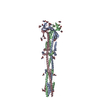
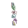
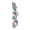
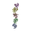
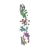
| Title | Tectonic conformational changes of a coronavirus spike glycoprotein promote membrane fusion | ||||||
 Components Components | Spike glycoprotein | ||||||
 Keywords Keywords | VIRAL PROTEIN / Coronavirus / membrane fusion / MHV / SARS / MERS | ||||||
| Function / homology |  Function and homology information Function and homology informationhost cell Golgi apparatus / host cell endoplasmic reticulum-Golgi intermediate compartment membrane / receptor-mediated virion attachment to host cell / endocytosis involved in viral entry into host cell / fusion of virus membrane with host plasma membrane / fusion of virus membrane with host endosome membrane / viral envelope / host cell plasma membrane / virion membrane / membrane / identical protein binding Similarity search - Function | ||||||
| Biological species |  Murine coronavirus Murine coronavirus | ||||||
| Method | ELECTRON MICROSCOPY / single particle reconstruction / cryo EM / Resolution: 4.1 Å | ||||||
 Authors Authors | Walls, A.C. / Tortorici, M.A. / Snijder, J. / Xiong, X. / Bosch, B.J. / Rey, F.A. / Veesler, D. | ||||||
| Funding support |  United States, 1items United States, 1items
| ||||||
 Citation Citation | Journal: Proc Natl Acad Sci U S A / Year: 2017 Title: Tectonic conformational changes of a coronavirus spike glycoprotein promote membrane fusion. Authors: Alexandra C Walls / M Alejandra Tortorici / Joost Snijder / Xiaoli Xiong / Berend-Jan Bosch / Felix A Rey / David Veesler /   Abstract: The tremendous pandemic potential of coronaviruses was demonstrated twice in the past few decades by two global outbreaks of deadly pneumonia. The coronavirus spike (S) glycoprotein initiates ...The tremendous pandemic potential of coronaviruses was demonstrated twice in the past few decades by two global outbreaks of deadly pneumonia. The coronavirus spike (S) glycoprotein initiates infection by promoting fusion of the viral and cellular membranes through conformational changes that remain largely uncharacterized. Here we report the cryoEM structure of a coronavirus S glycoprotein in the postfusion state, showing large-scale secondary, tertiary, and quaternary rearrangements compared with the prefusion trimer and rationalizing the free-energy landscape of this conformational machine. We also biochemically characterized the molecular events associated with refolding of the metastable prefusion S glycoprotein to the postfusion conformation using limited proteolysis, mass spectrometry, and single-particle EM. The observed similarity between postfusion coronavirus S and paramyxovirus F structures demonstrates that a conserved refolding trajectory mediates entry of these viruses and supports the evolutionary relatedness of their fusion subunits. Finally, our data provide a structural framework for understanding the mode of neutralization of antibodies targeting the fusion machinery and for engineering next-generation subunit vaccines or inhibitors against this medically important virus family. | ||||||
| History |
|
- Structure visualization
| Movie |
Movie viewer |
|---|---|
| Structure viewer | Molecule: MolmilJmol/JSmol |
- Downloads & links
-Download
| PDBx/mmCIF format | 6b3o.cif.gz | 216 KB | Display | PDBx/mmCIF format |
|---|---|---|---|---|
| PDB format | pdb6b3o.ent.gz | 167.2 KB | Display | PDB format |
| PDBx/mmJSON format | 6b3o.json.gz | Tree view | PDBx/mmJSON format | |
| Others |  Other downloads Other downloads |
-Validation report
| Arichive directory | https://data.pdbj.org/pub/pdb/validation_reports/b3/6b3oftp://data.pdbj.org/pub/pdb/validation_reports/b3/6b3o | HTTPS FTP |
|---|
-Related structure data
| Related structure data |  7040MC M: map data used to model this data C: citing same article ( |
|---|---|
| Similar structure data |


-Links
 PDBj
PDBj- Assembly
| Deposited unit | 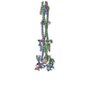
|
|---|---|
| 1 |
|
-Components
| #1: Protein | Mass: 66199.094 Da / Num. of mol.: 3 / Fragment: residues 718-1252 Source method: isolated from a genetically manipulated source Source: (gene. exp.) Murine coronavirus / Strain: A59 / Gene: S, 3 / Production host:  Has protein modification | Y | Sequence details | The authors state that all the differences between the sequence provided and the sequence database ...The authors state that all the differences between the sequence provided and the sequence database reference are accounted for by modifications that they made to their construct: residues 1253-1254: BiP secretion signal, residues 1253-1254: linker, residues 1255-1284: GCN4 trimerization motif, residues 1285-1290: Thrombin cleavage site, residues 1291-1300: Strep-tag | |
|---|
-Experimental details
-Experiment
| Experiment | Method: ELECTRON MICROSCOPY |
|---|---|
| EM experiment | Aggregation state: PARTICLE / 3D reconstruction method: single particle reconstruction |
- Sample preparation
| Component | Name: Mouse hepatitis virus spike glycoprotein (S2 subunit) in the postfusion conformation Type: COMPLEX / Entity ID: all / Source: RECOMBINANT |
|---|---|
| Molecular weight | Value: 0.18 MDa / Experimental value: NO |
| Source (natural) | Organism: Murine hepatitis virus |
| Source (recombinant) | Organism: |
| Buffer solution | pH: 7.5 |
| Specimen | Embedding applied: NO / Shadowing applied: NO / Staining applied: NO / Vitrification applied: YES |
| Specimen support | Grid material: COPPER / Grid mesh size: 400 divisions/in. / Grid type: C-flat |
| Vitrification | Cryogen name: ETHANE |
- Electron microscopy imaging
| Experimental equipment |  Model: Titan Krios / Image courtesy: FEI Company |
|---|---|
| Microscopy | Model: FEI TITAN KRIOS |
| Electron gun | Electron source:  FIELD EMISSION GUN / Accelerating voltage: 300 kV / Illumination mode: FLOOD BEAM FIELD EMISSION GUN / Accelerating voltage: 300 kV / Illumination mode: FLOOD BEAM |
| Electron lens | Mode: BRIGHT FIELD |
| Image recording | Electron dose: 60 e/Å2 / Detector mode: COUNTING / Film or detector model: GATAN K2 SUMMIT (4k x 4k) |
- Processing
| EM software |
| ||||||||||||||||||||||||||||||||
|---|---|---|---|---|---|---|---|---|---|---|---|---|---|---|---|---|---|---|---|---|---|---|---|---|---|---|---|---|---|---|---|---|---|
| CTF correction | Type: PHASE FLIPPING AND AMPLITUDE CORRECTION | ||||||||||||||||||||||||||||||||
| Symmetry | Point symmetry: C3 (3 fold cyclic) | ||||||||||||||||||||||||||||||||
| 3D reconstruction | Resolution: 4.1 Å / Resolution method: FSC 0.143 CUT-OFF / Num. of particles: 106000 / Symmetry type: POINT | ||||||||||||||||||||||||||||||||
| Atomic model building | Protocol: AB INITIO MODEL / Space: REAL |
