 Movie
Movie Controller

[English] 日本語
 Yorodumi
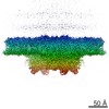
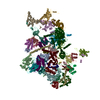
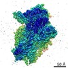
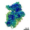
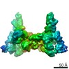
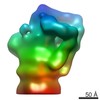
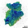
- PDB-6az1: Cryo-EM structure of the small subunit of Leishmania ribosome bou... -
+ Open data

- Basic information
| Entry | Database: PDB / ID: 6az1 | ||||||||||||||||||||||||||||||||||||
|---|---|---|---|---|---|---|---|---|---|---|---|---|---|---|---|---|---|---|---|---|---|---|---|---|---|---|---|---|---|---|---|---|---|---|---|---|---|
| Title | Cryo-EM structure of the small subunit of Leishmania ribosome bound to paromomycin | ||||||||||||||||||||||||||||||||||||
 Components Components |
| ||||||||||||||||||||||||||||||||||||
 Keywords Keywords | RIBOSOME/ANTIBIOTIC / Leishmania donovani / ribosome / aminoglycoside / paromomycin / RIBOSOME-ANTIBIOTIC complex | ||||||||||||||||||||||||||||||||||||
| Function / homology |  Function and homology information Function and homology information90S preribosome / translation regulator activity / maturation of SSU-rRNA from tricistronic rRNA transcript (SSU-rRNA, 5.8S rRNA, LSU-rRNA) / maturation of SSU-rRNA / small-subunit processome / rRNA processing / ribosomal small subunit assembly / ribosome binding / ribosomal small subunit biogenesis / small ribosomal subunit ...90S preribosome / translation regulator activity / maturation of SSU-rRNA from tricistronic rRNA transcript (SSU-rRNA, 5.8S rRNA, LSU-rRNA) / maturation of SSU-rRNA / small-subunit processome / rRNA processing / ribosomal small subunit assembly / ribosome binding / ribosomal small subunit biogenesis / small ribosomal subunit / small ribosomal subunit rRNA binding / cytosolic small ribosomal subunit / cytoplasmic translation / rRNA binding / structural constituent of ribosome / ribosome / translation / ribonucleoprotein complex / mRNA binding / nucleolus / RNA binding / zinc ion binding / nucleus / cytoplasm / cytosol Similarity search - Function | ||||||||||||||||||||||||||||||||||||
| Biological species |  Leishmania donovani (eukaryote) Leishmania donovani (eukaryote) | ||||||||||||||||||||||||||||||||||||
| Method | ELECTRON MICROSCOPY / single particle reconstruction / cryo EM / Resolution: 2.7 Å | ||||||||||||||||||||||||||||||||||||
 Authors Authors | Shalev-Benami, M. / Zhang, Y. / Rozenberg, H. / Matzov, D. / Zimmerman, E. / Bashan, A. / Jaffe, C.L. / Yonath, A. / Skiniotis, G. | ||||||||||||||||||||||||||||||||||||
 Citation Citation | Journal: Nat Commun / Year: 2017 Title: Atomic resolution snapshot of Leishmania ribosome inhibition by the aminoglycoside paromomycin. Authors: Moran Shalev-Benami / Yan Zhang / Haim Rozenberg / Yuko Nobe / Masato Taoka / Donna Matzov / Ella Zimmerman / Anat Bashan / Toshiaki Isobe / Charles L Jaffe / Ada Yonath / Georgios Skiniotis /    Abstract: Leishmania is a single-celled eukaryotic parasite afflicting millions of humans worldwide, with current therapies limited to a poor selection of drugs that mostly target elements in the parasite's ...Leishmania is a single-celled eukaryotic parasite afflicting millions of humans worldwide, with current therapies limited to a poor selection of drugs that mostly target elements in the parasite's cell envelope. Here we determined the atomic resolution electron cryo-microscopy (cryo-EM) structure of the Leishmania ribosome in complex with paromomycin (PAR), a highly potent compound recently approved for treatment of the fatal visceral leishmaniasis (VL). The structure reveals the mechanism by which the drug induces its deleterious effects on the parasite. We further show that PAR interferes with several aspects of cytosolic translation, thus highlighting the cytosolic rather than the mitochondrial ribosome as the primary drug target. The results also highlight unique as well as conserved elements in the PAR-binding pocket that can serve as hotspots for the development of novel therapeutics. | ||||||||||||||||||||||||||||||||||||
| History |
|
- Structure visualization
| Movie |
Movie viewer |
|---|---|
| Structure viewer | Molecule: MolmilJmol/JSmol |
- Downloads & links
-Download
| PDBx/mmCIF format | 6az1.cif.gz | 1.8 MB | Display | PDBx/mmCIF format |
|---|---|---|---|---|
| PDB format | pdb6az1.ent.gz | 1.4 MB | Display | PDB format |
| PDBx/mmJSON format | 6az1.json.gz | Tree view | PDBx/mmJSON format | |
| Others |  Other downloads Other downloads |
-Validation report
| Arichive directory | https://data.pdbj.org/pub/pdb/validation_reports/az/6az1ftp://data.pdbj.org/pub/pdb/validation_reports/az/6az1 | HTTPS FTP |
|---|
-Related structure data
| Related structure data | 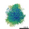 7024MC  7025C 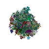 6az3C M: map data used to model this data C: citing same article ( |
|---|---|
| Similar structure data |

-Links
 PDBj
PDBj


- Assembly
| Deposited unit | 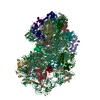
|
|---|---|
| 1 |
|
-Components
+Ribosomal protein ... , 32 types, 32 molecules ABCDEFGHIJKLMNOPQRSTUVWXYZabcdef
-Protein , 1 types, 1 molecules g
| #33: Protein | Mass: 34459.523 Da / Num. of mol.: 1 / Source method: isolated from a natural source / Source: (natural) Leishmania donovani (eukaryote) / References: UniProt: Q9BIJ5 |
|---|
-RNA chain , 5 types, 5 molecules 12345
| #34: RNA chain | Mass: 710287.438 Da / Num. of mol.: 1 / Source method: isolated from a natural source / Source: (natural) Leishmania donovani (eukaryote) / References: GenBank: 322500086 |
|---|---|
| #35: RNA chain | Mass: 24599.748 Da / Num. of mol.: 1 / Source method: obtained synthetically / Source: (synth.) |
| #36: RNA chain | Mass: 24786.785 Da / Num. of mol.: 1 / Source method: isolated from a natural source / Source: (natural) |
| #37: RNA chain | Mass: 24485.539 Da / Num. of mol.: 1 / Source method: isolated from a natural source / Source: (natural) |
| #38: RNA chain | Mass: 4085.501 Da / Num. of mol.: 1 / Source method: obtained synthetically / Source: (synth.) Leishmania donovani (eukaryote) |
-Non-polymers , 3 types, 292 molecules 
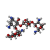

| #39: Chemical | ChemComp-MG /  Mass: 24.305 Da / Num. of mol.: 24 / Source method: obtained synthetically / Formula: Mg Mass: 24.305 Da / Num. of mol.: 24 / Source method: obtained synthetically / Formula: Mg#40: Chemical | ChemComp-PAR /  Mass: 615.628 Da / Num. of mol.: 6 / Source method: obtained synthetically / Formula: C23H45N5O14 / Comment: medication*YM Mass: 615.628 Da / Num. of mol.: 6 / Source method: obtained synthetically / Formula: C23H45N5O14 / Comment: medication*YM#41: Water | ChemComp-HOH / | Mass: 18.015 Da / Num. of mol.: 262 / Source method: isolated from a natural source / Formula: H2O |
|---|
-Details
| Has protein modification | Y |
|---|
-Experimental details
-Experiment
| Experiment | Method: ELECTRON MICROSCOPY |
|---|---|
| EM experiment | Aggregation state: PARTICLE / 3D reconstruction method: single particle reconstruction |
- Sample preparation
| Component | Name: Leishmania donovani 91S ribosome SSU / Type: RIBOSOME / Entity ID: #1-#38 / Source: NATURAL |
|---|---|
| Molecular weight | Experimental value: NO |
| Source (natural) | Organism: Leishmania donovani (eukaryote) |
| Buffer solution | pH: 7.6 |
| Specimen | Embedding applied: NO / Shadowing applied: NO / Staining applied: NO / Vitrification applied: YES |
| Vitrification | Cryogen name: ETHANE |
- Electron microscopy imaging
| Experimental equipment |  Model: Titan Krios / Image courtesy: FEI Company |
|---|---|
| Microscopy | Model: FEI TITAN KRIOS |
| Electron gun | Electron source:  FIELD EMISSION GUN / Accelerating voltage: 300 kV / Illumination mode: FLOOD BEAM FIELD EMISSION GUN / Accelerating voltage: 300 kV / Illumination mode: FLOOD BEAM |
| Electron lens | Mode: BRIGHT FIELD |
| Image recording | Electron dose: 1 e/Å2 / Film or detector model: GATAN K2 SUMMIT (4k x 4k) |
- Processing
| Software | Name: PHENIX / Version: dev-2686_1692: / Classification: refinement | ||||||||||||||||||||||||
|---|---|---|---|---|---|---|---|---|---|---|---|---|---|---|---|---|---|---|---|---|---|---|---|---|---|
| EM software | Name: PHENIX / Category: model refinement | ||||||||||||||||||||||||
| CTF correction | Type: PHASE FLIPPING AND AMPLITUDE CORRECTION | ||||||||||||||||||||||||
| 3D reconstruction | Resolution: 2.7 Å / Resolution method: FSC 0.143 CUT-OFF / Num. of particles: 141028 / Symmetry type: POINT | ||||||||||||||||||||||||
| Refine LS restraints |
|
