 Movie
Movie Controller

[English] 日本語
 Yorodumi
- PDB-6zgb: glutamate transporter homologue Glttk in complex with a photo cag... -
+ Open data

- Basic information
| Entry | Database: PDB / ID: 6zgb | ||||||
|---|---|---|---|---|---|---|---|
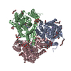
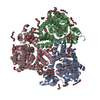
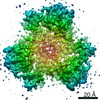
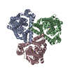
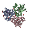
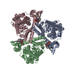
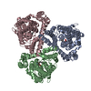
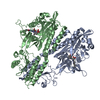
| Title | glutamate transporter homologue Glttk in complex with a photo cage compound | ||||||
 Components Components | Proton/glutamate symporter, SDF family | ||||||
 Keywords Keywords | TRANSPORT PROTEIN / glutamate transporter / photo cage / membrane protein | ||||||
| Function / homology |  Function and homology information Function and homology informationdicarboxylic acid transport / symporter activity / membrane / plasma membrane Similarity search - Function | ||||||
| Biological species |   Thermococcus kodakarensis (archaea) Thermococcus kodakarensis (archaea) | ||||||
| Method |  X-RAY DIFFRACTION / SYNCHROTRON / MOLECULAR REPLACEMENT / Resolution: 3.2 Å X-RAY DIFFRACTION / SYNCHROTRON / MOLECULAR REPLACEMENT / Resolution: 3.2 Å | ||||||
 Authors Authors | Arkhipova, V. / Slotboom, D.J. / Guskov, A. | ||||||
 Citation Citation | Journal: J.Am.Chem.Soc. / Year: 2021 Title: Structural Aspects of Photopharmacology: Insight into the Binding of Photoswitchable and Photocaged Inhibitors to the Glutamate Transporter Homologue. Authors: Arkhipova, V. / Fu, H. / Hoorens, M.W.H. / Trinco, G. / Lameijer, L.N. / Marin, E. / Feringa, B.L. / Poelarends, G.J. / Szymanski, W. / Slotboom, D.J. / Guskov, A. | ||||||
| History |
|
- Structure visualization
| Structure viewer | Molecule: MolmilJmol/JSmol |
|---|
- Downloads & links
-Download
| PDBx/mmCIF format | 6zgb.cif.gz | 493.2 KB | Display | PDBx/mmCIF format |
|---|---|---|---|---|
| PDB format | pdb6zgb.ent.gz | 414 KB | Display | PDB format |
| PDBx/mmJSON format | 6zgb.json.gz | Tree view | PDBx/mmJSON format | |
| Others |  Other downloads Other downloads |
-Validation report
| Arichive directory | https://data.pdbj.org/pub/pdb/validation_reports/zg/6zgbftp://data.pdbj.org/pub/pdb/validation_reports/zg/6zgb | HTTPS FTP |
|---|
-Related structure data
| Related structure data |  6zl4C  6zlhC 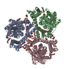 5dwyS S: Starting model for refinement C: citing same article ( |
|---|---|
| Similar structure data |

-Links
 PDBj
PDBj
- Assembly
| Deposited unit | 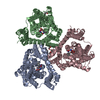
| |||||||||||||||||||||||||||||||||||||||||||||||||||||||||||||||||||||||||||||||||||||
|---|---|---|---|---|---|---|---|---|---|---|---|---|---|---|---|---|---|---|---|---|---|---|---|---|---|---|---|---|---|---|---|---|---|---|---|---|---|---|---|---|---|---|---|---|---|---|---|---|---|---|---|---|---|---|---|---|---|---|---|---|---|---|---|---|---|---|---|---|---|---|---|---|---|---|---|---|---|---|---|---|---|---|---|---|---|---|
| 1 |
| |||||||||||||||||||||||||||||||||||||||||||||||||||||||||||||||||||||||||||||||||||||
| Unit cell |
| |||||||||||||||||||||||||||||||||||||||||||||||||||||||||||||||||||||||||||||||||||||
| Noncrystallographic symmetry (NCS) | NCS domain:
NCS domain segments: Ens-ID: 1
|