 Movie
Movie Controller

[English] 日本語
 Yorodumi
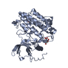
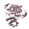
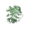
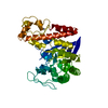
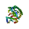
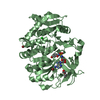
- PDB-6yg6: Crystal structure of MKK7 (MAP2K7) covalently bound with type-II ... -
+ Open data

- Basic information
| Entry | Database: PDB / ID: 6yg6 | ||||||
|---|---|---|---|---|---|---|---|
| Title | Crystal structure of MKK7 (MAP2K7) covalently bound with type-II inhibitor TL10-105 | ||||||
 Components Components | Dual specificity mitogen-activated protein kinase kinase 7 | ||||||
 Keywords Keywords | TRANSFERASE / kinase / kinase inhibitor / MKK7 / MEK7 / MAP2K7 / MAP2K / MEK / JNK signaling / Structural Genomics / Structural Genomics Consortium / SGC | ||||||
| Function / homology |  Function and homology information Function and homology informationJUN kinase kinase activity / regulation of motor neuron apoptotic process / mitogen-activated protein kinase kinase / response to osmotic stress / Fc-epsilon receptor signaling pathway / MAP kinase kinase activity / positive regulation of telomere maintenance / MAP kinase activity / Uptake and function of anthrax toxins / response to tumor necrosis factor ...JUN kinase kinase activity / regulation of motor neuron apoptotic process / mitogen-activated protein kinase kinase / response to osmotic stress / Fc-epsilon receptor signaling pathway / MAP kinase kinase activity / positive regulation of telomere maintenance / MAP kinase activity / Uptake and function of anthrax toxins / response to tumor necrosis factor / cellular response to interleukin-1 / stress-activated MAPK cascade / response to UV / JNK cascade / molecular function activator activity / JNK (c-Jun kinases) phosphorylation and activation mediated by activated human TAK1 / FCERI mediated MAPK activation / response to wounding / positive regulation of JNK cascade / cellular senescence / response to heat / cellular response to lipopolysaccharide / protein tyrosine kinase activity / protein phosphatase binding / Oxidative Stress Induced Senescence / positive regulation of ERK1 and ERK2 cascade / protein serine kinase activity / apoptotic process / protein kinase binding / positive regulation of DNA-templated transcription / magnesium ion binding / enzyme binding / signal transduction / ATP binding / nucleus / cytoplasm / cytosol Similarity search - Function | ||||||
| Biological species |  Homo sapiens (human) Homo sapiens (human) | ||||||
| Method |  X-RAY DIFFRACTION / SYNCHROTRON / MOLECULAR REPLACEMENT / Resolution: 2.15 Å X-RAY DIFFRACTION / SYNCHROTRON / MOLECULAR REPLACEMENT / Resolution: 2.15 Å | ||||||
 Authors Authors | Chaikuad, A. / Tan, L. / Wang, J. / Liang, Y. / Gray, N.S. / Knapp, S. / Structural Genomics Consortium (SGC) | ||||||
 Citation Citation | Journal: Cell Chem Biol / Year: 2020 Title: Catalytic Domain Plasticity of MKK7 Reveals Structural Mechanisms of Allosteric Activation and Diverse Targeting Opportunities. Authors: Schroder, M. / Tan, L. / Wang, J. / Liang, Y. / Gray, N.S. / Knapp, S. / Chaikuad, A. | ||||||
| History |
|
- Structure visualization
| Structure viewer | Molecule: MolmilJmol/JSmol |
|---|
- Downloads & links
-Download
| PDBx/mmCIF format | 6yg6.cif.gz | 249.7 KB | Display | PDBx/mmCIF format |
|---|---|---|---|---|
| PDB format | pdb6yg6.ent.gz | 200.3 KB | Display | PDB format |
| PDBx/mmJSON format | 6yg6.json.gz | Tree view | PDBx/mmJSON format | |
| Others |  Other downloads Other downloads |
-Validation report
| Arichive directory | https://data.pdbj.org/pub/pdb/validation_reports/yg/6yg6ftp://data.pdbj.org/pub/pdb/validation_reports/yg/6yg6 | HTTPS FTP |
|---|
-Related structure data
| Related structure data |  6yfzC  6yg0C  6yg1C 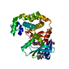 6yg2C  6yg3C  6yg4C  6yg5C  6yg7C 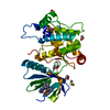 6yz4C 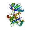 2dylS S: Starting model for refinement C: citing same article ( |
|---|---|
| Similar structure data |


-Links
 PDBj
PDBj


- Assembly
| Deposited unit | 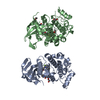
| ||||||||||||||||||
|---|---|---|---|---|---|---|---|---|---|---|---|---|---|---|---|---|---|---|---|
| 1 | 
| ||||||||||||||||||
| 2 | 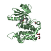
| ||||||||||||||||||
| Unit cell |
| ||||||||||||||||||
| Noncrystallographic symmetry (NCS) | NCS domain:
NCS domain segments: Component-ID: _ / Ens-ID: 1 / Beg auth comp-ID: GLY / Beg label comp-ID: GLY / End auth comp-ID: SER / End label comp-ID: SER / Refine code: _ / Auth seq-ID: 120 - 419 / Label seq-ID: 6 - 305
|
-Components
| #1: Protein | Mass: 34986.613 Da / Num. of mol.: 2 Source method: isolated from a genetically manipulated source Source: (gene. exp.) Homo sapiens (human) / Gene: MAP2K7, JNKK2, MEK7, MKK7, PRKMK7, SKK4 / Plasmid: pNIC28-Bsa4 / Production host:  References: UniProt: O14733, mitogen-activated protein kinase kinase #2: Chemical | ChemComp-EDO /   Mass: 62.068 Da / Num. of mol.: 4 / Source method: obtained synthetically / Formula: C2H6O2 Mass: 62.068 Da / Num. of mol.: 4 / Source method: obtained synthetically / Formula: C2H6O2#3: Chemical | 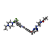  Mass: 639.711 Da / Num. of mol.: 2 / Source method: obtained synthetically / Formula: C33H40F3N7O3 / Feature type: SUBJECT OF INVESTIGATION Mass: 639.711 Da / Num. of mol.: 2 / Source method: obtained synthetically / Formula: C33H40F3N7O3 / Feature type: SUBJECT OF INVESTIGATION#4: Water | ChemComp-HOH / |  Mass: 18.015 Da / Num. of mol.: 163 / Source method: isolated from a natural source / Formula: H2O Mass: 18.015 Da / Num. of mol.: 163 / Source method: isolated from a natural source / Formula: H2OHas ligand of interest | Y | Has protein modification | Y | |
|---|
-Experimental details
-Experiment
| Experiment | Method: X-RAY DIFFRACTION / Number of used crystals: 1 |
|---|
- Sample preparation
| Crystal | Density Matthews: 2.33 Å3/Da / Density % sol: 47.3 % |
|---|---|
| Crystal grow | Temperature: 293.15 K / Method: vapor diffusion, sitting drop / pH: 8.2 Details: 19% PEG3350, 0.15 M ammonium acetate, 0.1 M tris, pH 8.2 |
-Data collection
| Diffraction | Mean temperature: 100 K / Serial crystal experiment: N | ||||||||||||||||||||||||||||||||||||||||||||||||||||||||||||||||||||||||||||||||||||||||||||||||||||||||||||||
|---|---|---|---|---|---|---|---|---|---|---|---|---|---|---|---|---|---|---|---|---|---|---|---|---|---|---|---|---|---|---|---|---|---|---|---|---|---|---|---|---|---|---|---|---|---|---|---|---|---|---|---|---|---|---|---|---|---|---|---|---|---|---|---|---|---|---|---|---|---|---|---|---|---|---|---|---|---|---|---|---|---|---|---|---|---|---|---|---|---|---|---|---|---|---|---|---|---|---|---|---|---|---|---|---|---|---|---|---|---|---|---|
| Diffraction source | Source: SYNCHROTRON / Site: Diamond  / Beamline: I02 / Wavelength: 0.97949 Å / Beamline: I02 / Wavelength: 0.97949 Å | ||||||||||||||||||||||||||||||||||||||||||||||||||||||||||||||||||||||||||||||||||||||||||||||||||||||||||||||
| Detector | Type: DECTRIS PILATUS3 6M / Detector: PIXEL / Date: Jul 23, 2015 | ||||||||||||||||||||||||||||||||||||||||||||||||||||||||||||||||||||||||||||||||||||||||||||||||||||||||||||||
| Radiation | Protocol: SINGLE WAVELENGTH / Monochromatic (M) / Laue (L): M / Scattering type: x-ray | ||||||||||||||||||||||||||||||||||||||||||||||||||||||||||||||||||||||||||||||||||||||||||||||||||||||||||||||
| Radiation wavelength | Wavelength: 0.97949 Å / Relative weight: 1 | ||||||||||||||||||||||||||||||||||||||||||||||||||||||||||||||||||||||||||||||||||||||||||||||||||||||||||||||
| Reflection | Resolution: 2.15→47.33 Å / Num. obs: 35192 / % possible obs: 99.9 % / Redundancy: 5.3 % / CC1/2: 0.996 / Rmerge(I) obs: 0.101 / Rpim(I) all: 0.05 / Rrim(I) all: 0.113 / Net I/av σ(I): 4.8 / Net I/σ(I): 10 | ||||||||||||||||||||||||||||||||||||||||||||||||||||||||||||||||||||||||||||||||||||||||||||||||||||||||||||||
| Reflection shell | Diffraction-ID: 1
|
- Processing
| Software |
| ||||||||||||||||||||||||||||||||||||||||||||||||||||||||||||||||||||||||||||||||||||||||||||||||||||||||||||||||||||||||||||||||||||||||||||||||||||||||||||||||||||||||||||||||||||||||||||||||||||||||
|---|---|---|---|---|---|---|---|---|---|---|---|---|---|---|---|---|---|---|---|---|---|---|---|---|---|---|---|---|---|---|---|---|---|---|---|---|---|---|---|---|---|---|---|---|---|---|---|---|---|---|---|---|---|---|---|---|---|---|---|---|---|---|---|---|---|---|---|---|---|---|---|---|---|---|---|---|---|---|---|---|---|---|---|---|---|---|---|---|---|---|---|---|---|---|---|---|---|---|---|---|---|---|---|---|---|---|---|---|---|---|---|---|---|---|---|---|---|---|---|---|---|---|---|---|---|---|---|---|---|---|---|---|---|---|---|---|---|---|---|---|---|---|---|---|---|---|---|---|---|---|---|---|---|---|---|---|---|---|---|---|---|---|---|---|---|---|---|---|---|---|---|---|---|---|---|---|---|---|---|---|---|---|---|---|---|---|---|---|---|---|---|---|---|---|---|---|---|---|---|---|---|
| Refinement | Method to determine structure: MOLECULAR REPLACEMENT Starting model: 2dyl Resolution: 2.15→47.33 Å / Cor.coef. Fo:Fc: 0.954 / Cor.coef. Fo:Fc free: 0.942 / SU B: 14.242 / SU ML: 0.181 / SU R Cruickshank DPI: 0.2578 / Cross valid method: THROUGHOUT / σ(F): 0 / ESU R: 0.258 / ESU R Free: 0.198 Details: U VALUES : WITH TLS ADDED HYDROGENS HAVE BEEN ADDED IN THE RIDING POSITIONS
| ||||||||||||||||||||||||||||||||||||||||||||||||||||||||||||||||||||||||||||||||||||||||||||||||||||||||||||||||||||||||||||||||||||||||||||||||||||||||||||||||||||||||||||||||||||||||||||||||||||||||
| Solvent computation | Ion probe radii: 0.8 Å / Shrinkage radii: 0.8 Å / VDW probe radii: 1.2 Å | ||||||||||||||||||||||||||||||||||||||||||||||||||||||||||||||||||||||||||||||||||||||||||||||||||||||||||||||||||||||||||||||||||||||||||||||||||||||||||||||||||||||||||||||||||||||||||||||||||||||||
| Displacement parameters | Biso max: 164.97 Å2 / Biso mean: 62.631 Å2 / Biso min: 31.11 Å2
| ||||||||||||||||||||||||||||||||||||||||||||||||||||||||||||||||||||||||||||||||||||||||||||||||||||||||||||||||||||||||||||||||||||||||||||||||||||||||||||||||||||||||||||||||||||||||||||||||||||||||
| Refinement step | Cycle: final / Resolution: 2.15→47.33 Å
| ||||||||||||||||||||||||||||||||||||||||||||||||||||||||||||||||||||||||||||||||||||||||||||||||||||||||||||||||||||||||||||||||||||||||||||||||||||||||||||||||||||||||||||||||||||||||||||||||||||||||
| Refine LS restraints |
| ||||||||||||||||||||||||||||||||||||||||||||||||||||||||||||||||||||||||||||||||||||||||||||||||||||||||||||||||||||||||||||||||||||||||||||||||||||||||||||||||||||||||||||||||||||||||||||||||||||||||
| Refine LS restraints NCS | Ens-ID: 1 / Number: 34370 / Refine-ID: X-RAY DIFFRACTION / Type: interatomic distance / Rms dev position: 0.09 Å / Weight position: 0.05
| ||||||||||||||||||||||||||||||||||||||||||||||||||||||||||||||||||||||||||||||||||||||||||||||||||||||||||||||||||||||||||||||||||||||||||||||||||||||||||||||||||||||||||||||||||||||||||||||||||||||||
| LS refinement shell | Resolution: 2.15→2.206 Å / Rfactor Rfree error: 0 / Total num. of bins used: 20
| ||||||||||||||||||||||||||||||||||||||||||||||||||||||||||||||||||||||||||||||||||||||||||||||||||||||||||||||||||||||||||||||||||||||||||||||||||||||||||||||||||||||||||||||||||||||||||||||||||||||||
| Refinement TLS params. | Method: refined / Refine-ID: X-RAY DIFFRACTION
| ||||||||||||||||||||||||||||||||||||||||||||||||||||||||||||||||||||||||||||||||||||||||||||||||||||||||||||||||||||||||||||||||||||||||||||||||||||||||||||||||||||||||||||||||||||||||||||||||||||||||
| Refinement TLS group |
|
