 Movie
Movie Controller

[English] 日本語
 Yorodumi
- PDB-6xsp: Crystal structure of E.coli DsbA in complex with 2-(2,6-bis(3-met... -
+ Open data

- Basic information
| Entry | Database: PDB / ID: 6xsp | ||||||
|---|---|---|---|---|---|---|---|
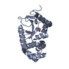
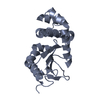
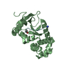
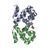
| Title | Crystal structure of E.coli DsbA in complex with 2-(2,6-bis(3-methoxyphenyl)benzofuran-3-yl)acetic acid | ||||||
 Components Components | Thiol:disulfide interchange protein DsbA | ||||||
 Keywords Keywords | OXIDOREDUCTASE/INHIBITOR / Inhibitor / complex / disulfide oxidoreductase / fragments / OXIDOREDUCTASE-OXIDOREDUCTASE INHIBITOR complex / OXIDOREDUCTASE / OXIDOREDUCTASE-INHIBITOR complex | ||||||
| Function / homology |  Function and homology information Function and homology informationSecretion of toxins / protein disulfide isomerase activity / cellular response to antibiotic / protein-disulfide reductase activity / outer membrane-bounded periplasmic space Similarity search - Function | ||||||
| Biological species |  | ||||||
| Method |  X-RAY DIFFRACTION / SYNCHROTRON / MOLECULAR REPLACEMENT / Resolution: 2.3 Å X-RAY DIFFRACTION / SYNCHROTRON / MOLECULAR REPLACEMENT / Resolution: 2.3 Å | ||||||
 Authors Authors | Wang, G. / Heras, B. | ||||||
| Funding support |  Australia, 1items Australia, 1items
| ||||||
 Citation Citation | Journal: Bioorg.Med.Chem. / Year: 2021 Title: Elaboration of a benzofuran scaffold and evaluation of binding affinity and inhibition of Escherichia coli DsbA: A fragment-based drug design approach to novel antivirulence compounds. Authors: Duncan, L.F. / Wang, G. / Ilyichova, O.V. / Dhouib, R. / Totsika, M. / Scanlon, M.J. / Heras, B. / Abbott, B.M. | ||||||
| History |
|
- Structure visualization
| Structure viewer | Molecule: MolmilJmol/JSmol |
|---|
- Downloads & links
-Download
| PDBx/mmCIF format | 6xsp.cif.gz | 92.6 KB | Display | PDBx/mmCIF format |
|---|---|---|---|---|
| PDB format | pdb6xsp.ent.gz | 68.5 KB | Display | PDB format |
| PDBx/mmJSON format | 6xsp.json.gz | Tree view | PDBx/mmJSON format | |
| Others |  Other downloads Other downloads |
-Validation report
| Arichive directory | https://data.pdbj.org/pub/pdb/validation_reports/xs/6xspftp://data.pdbj.org/pub/pdb/validation_reports/xs/6xsp | HTTPS FTP |
|---|
-Related structure data
| Related structure data |  6xsqC  6xt3C  7l76C  7l7cC  7lhpC  1fvkS S: Starting model for refinement C: citing same article ( |
|---|---|
| Similar structure data |


-Links
 PDBj
PDBj

- Assembly
| Deposited unit | 
| ||||||||
|---|---|---|---|---|---|---|---|---|---|
| 1 | 
| ||||||||
| 2 | 
| ||||||||
| Unit cell |
| ||||||||
| Components on special symmetry positions |
|
-Components
| #1: Protein | Mass: 21155.025 Da / Num. of mol.: 2 Source method: isolated from a genetically manipulated source Source: (gene. exp.) Strain: K12 / Gene: dsbA, dsf, ppfA, b3860, JW3832 / Production host: #2: Chemical | ChemComp-VCY / [ |   Mass: 388.413 Da / Num. of mol.: 1 / Source method: obtained synthetically / Formula: C24H20O5 / Feature type: SUBJECT OF INVESTIGATION Mass: 388.413 Da / Num. of mol.: 1 / Source method: obtained synthetically / Formula: C24H20O5 / Feature type: SUBJECT OF INVESTIGATION#3: Chemical | ChemComp-CU / |   Mass: 63.546 Da / Num. of mol.: 1 / Source method: obtained synthetically / Formula: Cu Mass: 63.546 Da / Num. of mol.: 1 / Source method: obtained synthetically / Formula: Cu#4: Water | ChemComp-HOH / |  Mass: 18.015 Da / Num. of mol.: 205 / Source method: isolated from a natural source / Formula: H2O Mass: 18.015 Da / Num. of mol.: 205 / Source method: isolated from a natural source / Formula: H2OHas ligand of interest | Y | Has protein modification | Y | |
|---|
-Experimental details
-Experiment
| Experiment | Method: X-RAY DIFFRACTION / Number of used crystals: 1 |
|---|
- Sample preparation
| Crystal | Density Matthews: 2.67 Å3/Da / Density % sol: 53.85 % |
|---|---|
| Crystal grow | Temperature: 293 K / Method: vapor diffusion, hanging drop Details: 11-13% PEG 8000, 5-7.5% glycerol, 1 mM copper(II) chloride, 100 mM sodium cacodylate |
-Data collection
| Diffraction | Mean temperature: 100 K / Serial crystal experiment: N | ||||||||||||||||||||||||||||||
|---|---|---|---|---|---|---|---|---|---|---|---|---|---|---|---|---|---|---|---|---|---|---|---|---|---|---|---|---|---|---|---|
| Diffraction source | Source: SYNCHROTRON / Site: Australian Synchrotron / Beamline: MX1 / Wavelength: 0.9537 Å | ||||||||||||||||||||||||||||||
| Detector | Type: ADSC QUANTUM 210r / Detector: CCD / Date: Dec 6, 2016 | ||||||||||||||||||||||||||||||
| Radiation | Protocol: SINGLE WAVELENGTH / Monochromatic (M) / Laue (L): M / Scattering type: x-ray | ||||||||||||||||||||||||||||||
| Radiation wavelength | Wavelength: 0.9537 Å / Relative weight: 1 | ||||||||||||||||||||||||||||||
| Reflection | Resolution: 2.3→31.91 Å / Num. obs: 19927 / % possible obs: 99.9 % / Redundancy: 4 % / CC1/2: 0.997 / Rmerge(I) obs: 0.062 / Rpim(I) all: 0.035 / Rrim(I) all: 0.072 / Net I/σ(I): 13.8 / Num. measured all: 80257 / Scaling rejects: 331 | ||||||||||||||||||||||||||||||
| Reflection shell | Diffraction-ID: 1
|
- Processing
| Software |
| ||||||||||||||||||||||||||||||||||||||||||||||||
|---|---|---|---|---|---|---|---|---|---|---|---|---|---|---|---|---|---|---|---|---|---|---|---|---|---|---|---|---|---|---|---|---|---|---|---|---|---|---|---|---|---|---|---|---|---|---|---|---|---|
| Refinement | Method to determine structure: MOLECULAR REPLACEMENT Starting model: 1FVK Resolution: 2.3→31.91 Å / SU ML: 0.32 / Cross valid method: THROUGHOUT / σ(F): 1.35 / Phase error: 26.48 / Stereochemistry target values: ML
| ||||||||||||||||||||||||||||||||||||||||||||||||
| Solvent computation | Shrinkage radii: 0.9 Å / VDW probe radii: 1.11 Å / Solvent model: FLAT BULK SOLVENT MODEL | ||||||||||||||||||||||||||||||||||||||||||||||||
| Displacement parameters | Biso max: 89.23 Å2 / Biso mean: 34.8949 Å2 / Biso min: 11.69 Å2 | ||||||||||||||||||||||||||||||||||||||||||||||||
| Refinement step | Cycle: final / Resolution: 2.3→31.91 Å
| ||||||||||||||||||||||||||||||||||||||||||||||||
| Refine LS restraints |
| ||||||||||||||||||||||||||||||||||||||||||||||||
| LS refinement shell | Refine-ID: X-RAY DIFFRACTION / Rfactor Rfree error: 0 / Total num. of bins used: 7 / % reflection obs: 100 %
|
