National Institutes of Health/National Institute of General Medical Sciences (NIH/NIGMS)
R35GM122510
United States
National Institutes of Health/National Institute of General Medical Sciences (NIH/NIGMS)
T32GM080186
United States
Citation
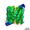
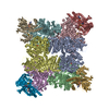
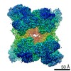
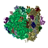
Journal: Proc Natl Acad Sci U S A / Year: 2020 Title: Atomic structure of the flagellar filament reveals how ε Proteobacteria escaped Toll-like receptor 5 surveillance. Authors: Mark A B Kreutzberger / Cheryl Ewing / Frederic Poly / Fengbin Wang / Edward H Egelman / Abstract: Vertebrates, from zebra fish to humans, have an innate immune recognition of many bacterial flagellins. This involves a conserved eight-amino acid epitope in flagellin recognized by the Toll-like ...Vertebrates, from zebra fish to humans, have an innate immune recognition of many bacterial flagellins. This involves a conserved eight-amino acid epitope in flagellin recognized by the Toll-like receptor 5 (TLR5). Several important human pathogens, such as and , have escaped TLR5 activation by mutations in this epitope. When such mutations were introduced into flagellin, motility was abolished. It was previously argued, using very low-resolution cryoelectron microscopy (cryo-EM), that accommodated these mutations by forming filaments with 7 protofilaments, rather than the 11 found in other bacteria. We have now determined the atomic structure of the G508A flagellar filament from a 3.5-Å-resolution cryo-EM reconstruction, and show that it has 11 protofilaments. The residues in the TLR5 epitope have reduced contacts with the adjacent subunit compared to other bacterial flagellar filament structures. The weakening of the subunit-subunit interface introduced by the mutations in the TLR5 epitope is compensated for by extensive interactions between the outer domains of the flagellin subunits. In other bacteria, these outer domains can be nearly absent or removed without affecting motility. Furthermore, we provide evidence for the stabilization of these outer domain interactions through glycosylation of key residues. These results explain the essential role of glycosylation in motility, and show how the outer domains have evolved to play a role not previously found in other bacteria.
History
Deposition
Jun 1, 2020
Deposition site: RCSB / Processing site: RCSB
Revision 1.0
Jul 8, 2020
Provider: repository / Type: Initial release
Revision 1.0
Jul 8, 2020
Data content type: EM metadata / Data content type: EM metadata / Provider: repository / Type: Initial release
Revision 1.0
Jul 8, 2020
Data content type: Image / Data content type: Image / Provider: repository / Type: Initial release
Revision 1.0
Jul 8, 2020
Data content type: Primary map / Data content type: Primary map / Provider: repository / Type: Initial release
Revision 1.0
Jul 8, 2020
Data content type: Image / Data content type: Image / Provider: repository / Type: Initial release
Revision 1.0
Jul 8, 2020
Data content type: Primary map / Data content type: Primary map / Provider: repository / Type: Initial release
Revision 1.0
Jul 8, 2020
Data content type: Image / Data content type: Image / Provider: repository / Type: Initial release
Revision 1.0
Jul 8, 2020
Data content type: Primary map / Data content type: Primary map / Provider: repository / Type: Initial release
Revision 1.0
Jul 8, 2020
Data content type: Image / Data content type: Image / Provider: repository / Type: Initial release
Revision 1.0
Jul 8, 2020
Data content type: Primary map / Data content type: Primary map / Provider: repository / Type: Initial release
Revision 1.0
Jul 8, 2020
Data content type: Image / Data content type: Image / Provider: repository / Type: Initial release
Revision 1.0
Jul 8, 2020
Data content type: Primary map / Data content type: Primary map / Provider: repository / Type: Initial release
Data content type: EM metadata / Data content type: EM metadata / EM metadata / Group: Data processing / Experimental summary / Data content type: EM metadata / EM metadata / Category: em_admin / em_software / Data content type: EM metadata / EM metadata / Item: _em_admin.last_update / _em_software.name
A: Flagellin A B: Flagellin A C: Flagellin A D: Flagellin A E: Flagellin A F: Flagellin A G: Flagellin A H: Flagellin A I: Flagellin A J: Flagellin A K: Flagellin A L: Flagellin A M: Flagellin A N: Flagellin A O: Flagellin A P: Flagellin A Q: Flagellin A R: Flagellin A S: Flagellin A T: Flagellin A U: Flagellin A V: Flagellin A hetero molecules
In the structure databanks used in Yorodumi, some data are registered as the other names, "COVID-19 virus" and "2019-nCoV". Here are the details of the virus and the list of structure data.
Jan 31, 2019. EMDB accession codes are about to change! (news from PDBe EMDB page)
EMDB accession codes are about to change! (news from PDBe EMDB page)
The allocation of 4 digits for EMDB accession codes will soon come to an end. Whilst these codes will remain in use, new EMDB accession codes will include an additional digit and will expand incrementally as the available range of codes is exhausted. The current 4-digit format prefixed with “EMD-” (i.e. EMD-XXXX) will advance to a 5-digit format (i.e. EMD-XXXXX), and so on. It is currently estimated that the 4-digit codes will be depleted around Spring 2019, at which point the 5-digit format will come into force.
The EM Navigator/Yorodumi systems omit the EMD- prefix.
Related info.:Q: What is EMD? / ID/Accession-code notation in Yorodumi/EM Navigator
Yorodumi is a browser for structure data from EMDB, PDB, SASBDB, etc.
This page is also the successor to EM Navigator detail page, and also detail information page/front-end page for Omokage search.
The word "yorodu" (or yorozu) is an old Japanese word meaning "ten thousand". "mi" (miru) is to see.
Related info.:EMDB / PDB / SASBDB / Comparison of 3 databanks / Yorodumi Search / Aug 31, 2016. New EM Navigator & Yorodumi / Yorodumi Papers / Jmol/JSmol / Function and homology information / Changes in new EM Navigator and Yorodumi
 Movie
Movie Controller


 Open data
 Basic information
 Components
 Keywords
 Function and homology information
 Campylobacter jejuni (Campylobacter)
 Authors
 United States, 2items
  Citation
 Structure visualization
 Downloads & links
 Other downloads
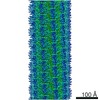

 PDBj
PDBj Assembly
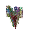

 Type: L-saccharide, alpha linking / Mass: 250.249 Da / Num. of mol.: 374 / Source method: obtained synthetically / Formula: C9H18N2O6 / Feature type: SUBJECT OF INVESTIGATION
 Sample preparation
 Electron microscopy imaging
 FIELD EMISSION GUN / Accelerating voltage: 300 kV / Illumination mode: FLOOD BEAM
 Processing