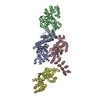
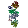
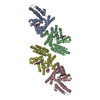
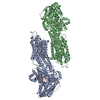
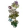
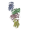
Journal: Nature / Year: 2020 Title: Structure of nevanimibe-bound tetrameric human ACAT1. Authors: Tao Long / Yingyuan Sun / Abdirahman Hassan / Xiaofeng Qi / Xiaochun Li / Abstract: Cholesterol is an essential component of mammalian cell membranes, constituting up to 50% of plasma membrane lipids. By contrast, it accounts for only 5% of lipids in the endoplasmic reticulum (ER). ...Cholesterol is an essential component of mammalian cell membranes, constituting up to 50% of plasma membrane lipids. By contrast, it accounts for only 5% of lipids in the endoplasmic reticulum (ER). The ER enzyme sterol O-acyltransferase 1 (also named acyl-coenzyme A:cholesterol acyltransferase, ACAT1) transfers a long-chain fatty acid to cholesterol to form cholesteryl esters that coalesce into cytosolic lipid droplets. Under conditions of cholesterol overload, ACAT1 maintains the low cholesterol concentration of the ER and thereby has an essential role in cholesterol homeostasis. ACAT1 has also been implicated in Alzheimer's disease, atherosclerosis and cancers. Here we report a cryo-electron microscopy structure of human ACAT1 in complex with nevanimibe, an inhibitor that is in clinical trials for the treatment of congenital adrenal hyperplasia. The ACAT1 holoenzyme is a tetramer that consists of two homodimers. Each monomer contains nine transmembrane helices (TMs), six of which (TM4-TM9) form a cavity that accommodates nevanimibe and an endogenous acyl-coenzyme A. This cavity also contains a histidine that has previously been identified as essential for catalytic activity. Our structural data and biochemical analyses provide a physical model to explain the process of cholesterol esterification, as well as details of the interaction between nevanimibe and ACAT1, which may help to accelerate the development of ACAT1 inhibitors to treat related diseases.
Resolution: 3.67 Å / Resolution method: FSC 0.143 CUT-OFF / Num. of particles: 263839 / Symmetry type: POINT
Refinement
Resolution: 3.67→274.89 Å / Cor.coef. Fo:Fc: 0.801 / SU B: 22.557 / SU ML: 0.327 / ESU R: 0.213 / Stereochemistry target values: MAXIMUM LIKELIHOOD / Details: HYDROGENS HAVE BEEN ADDED IN THE RIDING POSITIONS
Rfactor
Num. reflection
% reflection
Rwork
0.4387
-
-
obs
0.4387
880402
100 %
Solvent computation
Ion probe radii: 0.8 Å / Shrinkage radii: 0.8 Å / VDW probe radii: 1.2 Å / Solvent model: MASK
 Movie
Movie Controller


 Open data
 Basic information
 Components
 Keywords
 Function and homology information
 Homo sapiens (human)
 Authors
 Citation
 Structure visualization
 Downloads & links
 Other downloads
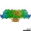


 PDBj
PDBj


 Assembly


 Mass: 282.461 Da / Num. of mol.: 4 / Source method: obtained synthetically / Formula: C18H34O2
 Mass: 386.654 Da / Num. of mol.: 10 / Source method: obtained synthetically / Formula: C27H46O
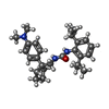
 Mass: 421.618 Da / Num. of mol.: 4 / Source method: obtained synthetically / Formula: C27H39N3O / Feature type: SUBJECT OF INVESTIGATION
 Mass: 767.534 Da / Num. of mol.: 2 / Source method: obtained synthetically / Formula: C21H36N7O16P3S
 Mass: 1031.980 Da / Num. of mol.: 2 / Source method: obtained synthetically / Formula: C39H68N7O17P3S / Feature type: SUBJECT OF INVESTIGATION
 Sample preparation
 Electron microscopy imaging
 FIELD EMISSION GUN / Accelerating voltage: 300 kV / Illumination mode: FLOOD BEAM
 Processing