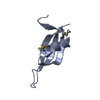
 Movie
Movie Controller

[English] 日本語
 Yorodumi
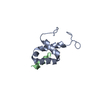
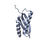
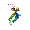
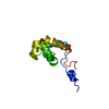
- PDB-6s6r: First crystal structure of parasitic PEX14 in complex with a frag... -
+ Open data

- Basic information
| Entry | Database: PDB / ID: 6s6r | ||||||
|---|---|---|---|---|---|---|---|
| Title | First crystal structure of parasitic PEX14 in complex with a fragment molecule 1H-indole-7-carboxylic acid | ||||||
 Components Components | Peroxin 14 | ||||||
 Keywords Keywords | SIGNALING PROTEIN / PEX14 / PPI / PEX14/PEX5 / Trypanosomes / FBDD | ||||||
| Function / homology |  Function and homology information Function and homology informationglycosome membrane / peroxisomal importomer complex / protein import into peroxisome matrix, docking / glycosome / protein targeting to vacuole / post-transcriptional regulation of gene expression / signaling receptor binding Similarity search - Function | ||||||
| Biological species |  | ||||||
| Method |  X-RAY DIFFRACTION / SYNCHROTRON / MOLECULAR REPLACEMENT / Resolution: 1.58000903163 Å X-RAY DIFFRACTION / SYNCHROTRON / MOLECULAR REPLACEMENT / Resolution: 1.58000903163 Å | ||||||
 Authors Authors | Hassaan, E. / Heine, A. / Klebe, G. | ||||||
| Funding support |  Germany, 1items Germany, 1items
| ||||||
 Citation Citation | Journal: To Be Published Title: First crystal structure of parasitic PEX14 in complex with a fragment molecule 1H-indole-7-carboxylic acid Authors: Hassaan, E. / Heine, A. / Klebe, G. | ||||||
| History |
|
- Structure visualization
| Structure viewer | Molecule: MolmilJmol/JSmol |
|---|
- Downloads & links
-Download
| PDBx/mmCIF format | 6s6r.cif.gz | 50.7 KB | Display | PDBx/mmCIF format |
|---|---|---|---|---|
| PDB format | pdb6s6r.ent.gz | 30 KB | Display | PDB format |
| PDBx/mmJSON format | 6s6r.json.gz | Tree view | PDBx/mmJSON format | |
| Others |  Other downloads Other downloads |
-Validation report
| Arichive directory | https://data.pdbj.org/pub/pdb/validation_reports/s6/6s6rftp://data.pdbj.org/pub/pdb/validation_reports/s6/6s6r | HTTPS FTP |
|---|
-Related structure data
| Similar structure data |
|---|


-Links
 PDBj
PDBj- Assembly
| Deposited unit | 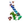
| ||||||||||
|---|---|---|---|---|---|---|---|---|---|---|---|
| 1 |
| ||||||||||
| Unit cell |
|
-Components
-Protein , 1 types, 1 molecules A
| #1: Protein | Mass: 7883.075 Da / Num. of mol.: 1 Source method: isolated from a genetically manipulated source Source: (gene. exp.)  |
|---|
-Non-polymers , 5 types, 48 molecules 
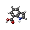


| #2: Chemical |  Mass: 96.063 Da / Num. of mol.: 3 / Source method: obtained synthetically / Formula: SO4 Mass: 96.063 Da / Num. of mol.: 3 / Source method: obtained synthetically / Formula: SO4#3: Chemical | ChemComp-KXQ / |  Mass: 161.157 Da / Num. of mol.: 1 / Source method: obtained synthetically / Formula: C9H7NO2 / Feature type: SUBJECT OF INVESTIGATION Mass: 161.157 Da / Num. of mol.: 1 / Source method: obtained synthetically / Formula: C9H7NO2 / Feature type: SUBJECT OF INVESTIGATION#4: Chemical | ChemComp-GOL / |  Mass: 92.094 Da / Num. of mol.: 1 / Source method: obtained synthetically / Formula: C3H8O3 Mass: 92.094 Da / Num. of mol.: 1 / Source method: obtained synthetically / Formula: C3H8O3#5: Chemical | ChemComp-DMS / |  Mass: 78.133 Da / Num. of mol.: 1 / Source method: obtained synthetically / Formula: C2H6OS / Comment: DMSO, precipitant*YM Mass: 78.133 Da / Num. of mol.: 1 / Source method: obtained synthetically / Formula: C2H6OS / Comment: DMSO, precipitant*YM#6: Water | ChemComp-HOH / | Mass: 18.015 Da / Num. of mol.: 42 / Source method: isolated from a natural source / Formula: H2O |
|---|
-Details
| Has ligand of interest | Y |
|---|
-Experimental details
-Experiment
| Experiment | Method: X-RAY DIFFRACTION / Number of used crystals: 1 |
|---|
- Sample preparation
| Crystal | Density Matthews: 2.41 Å3/Da / Density % sol: 48.9 % |
|---|---|
| Crystal grow | Temperature: 277 K / Method: vapor diffusion, sitting drop / Details: 0.2 M ammonium sulphate, 50% PEG 8000, 5% glycerol |
-Data collection
| Diffraction | Mean temperature: 100 K / Serial crystal experiment: N |
|---|---|
| Diffraction source | Source: SYNCHROTRON / Site: BESSY / Beamline: 14.1 / Wavelength: 0.9184 Å |
| Detector | Type: DECTRIS PILATUS 6M / Detector: PIXEL / Date: Nov 25, 2016 |
| Radiation | Protocol: SINGLE WAVELENGTH / Monochromatic (M) / Laue (L): M / Scattering type: x-ray |
| Radiation wavelength | Wavelength: 0.9184 Å / Relative weight: 1 |
| Reflection | Resolution: 1.58→43.306 Å / Num. obs: 10965 / % possible obs: 98.4 % / Redundancy: 8.22 % / Biso Wilson estimate: 18.9861182796 Å2 / CC1/2: 0.998 / Rrim(I) all: 0.074 / Net I/σ(I): 16.93 |
| Reflection shell | Resolution: 1.58→1.588 Å / Num. unique obs: 10965 |
- Processing
| Software |
| |||||||||||||||||||||||||||||||||||
|---|---|---|---|---|---|---|---|---|---|---|---|---|---|---|---|---|---|---|---|---|---|---|---|---|---|---|---|---|---|---|---|---|---|---|---|---|
| Refinement | Method to determine structure: MOLECULAR REPLACEMENT / Resolution: 1.58000903163→38.1843205977 Å / SU ML: 0.165444141052 / Cross valid method: FREE R-VALUE / σ(F): 1.37743608206 / Phase error: 23.5566591667
| |||||||||||||||||||||||||||||||||||
| Solvent computation | Shrinkage radii: 0.9 Å / VDW probe radii: 1.11 Å | |||||||||||||||||||||||||||||||||||
| Displacement parameters | Biso mean: 23.7840108874 Å2 | |||||||||||||||||||||||||||||||||||
| Refinement step | Cycle: LAST / Resolution: 1.58000903163→38.1843205977 Å
| |||||||||||||||||||||||||||||||||||
| Refine LS restraints |
| |||||||||||||||||||||||||||||||||||
| LS refinement shell | Refine-ID: X-RAY DIFFRACTION
|
