 Movie
Movie Controller

[English] 日本語
 Yorodumi
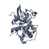
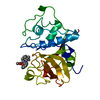
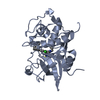
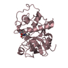
- PDB-6pad: Binding of chloromethyl ketone substrate analogues to crystalline... -
+ Open data

- Basic information
| Entry | Database: PDB / ID: 6pad | ||||||||||||
|---|---|---|---|---|---|---|---|---|---|---|---|---|---|
| Title | Binding of chloromethyl ketone substrate analogues to crystalline papain | ||||||||||||
 Components Components | PAPAIN | ||||||||||||
 Keywords Keywords | HYDROLASE/HYDROLASE INHIBITOR / SULFHYDRYL PROTEINASE / HYDROLASE-HYDROLASE INHIBITOR complex | ||||||||||||
| Function / homology |  Function and homology information Function and homology informationpapain / serpin family protein binding / cysteine-type peptidase activity / proteolysis Similarity search - Function | ||||||||||||
| Biological species |  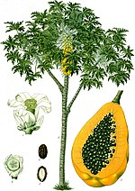 Carica papaya (papaya) Carica papaya (papaya) | ||||||||||||
| Method |  X-RAY DIFFRACTION / Resolution: 2.8 Å X-RAY DIFFRACTION / Resolution: 2.8 Å | ||||||||||||
 Authors Authors | Drenth, J. / Kalk, K.H. / Swen, H.M. | ||||||||||||
 Citation Citation | Journal: Biochemistry / Year: 1976 Title: Binding of chloromethyl ketone substrate analogues to crystalline papain. Authors: Drenth, J. / Kalk, K.H. / Swen, H.M. #1: Journal: Adv.Protein Chem. / Year: 1971Title: The Structure of Papain Authors: Drenth, J. / Jansonius, J.N. / Koekoek, R. / Wolthers, B.G. #2: Journal: Philos.Trans.R.Soc.London,Ser.B / Year: 1970Title: The Structure of the Papain Molecule Authors: Drenth, J. / Jansonius, J.N. / Koekoek, R. / Sluyterman, L.A.A. / Wolthers, B.G. #3: Journal: Nature / Year: 1968Title: Structure of Papain Authors: Drenth, J. / Jansonius, J.N. / Koekoek, R. / Swen, H.M. / Wolthers, B.G. | ||||||||||||
| History |
| ||||||||||||
| Remark 700 | SHEET THE SHEET SUBSTRUCTURE OF THIS MOLECULE IS DOUBLY BIFURCATED. IN ORDER TO REPRESENT THIS ...SHEET THE SHEET SUBSTRUCTURE OF THIS MOLECULE IS DOUBLY BIFURCATED. IN ORDER TO REPRESENT THIS FEATURE IN THE SHEET RECORDS BELOW, TWO SHEETS ARE DEFINED. STRANDS 4,5,6 OF S1A ARE IDENTICAL TO STRANDS 2,3,4 OF S1B. |
- Structure visualization
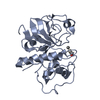
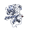
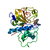
| Structure viewer | Molecule: MolmilJmol/JSmol |
|---|
- Downloads & links
-Download
| PDBx/mmCIF format | 6pad.cif.gz | 51.7 KB | Display | PDBx/mmCIF format |
|---|---|---|---|---|
| PDB format | pdb6pad.ent.gz | 36.3 KB | Display | PDB format |
| PDBx/mmJSON format | 6pad.json.gz | Tree view | PDBx/mmJSON format | |
| Others |  Other downloads Other downloads |
-Validation report
| Summary document | 6pad_validation.pdf.gz | 457.4 KB | Display | wwPDB validaton report |
|---|---|---|---|---|
| Full document | 6pad_full_validation.pdf.gz | 490.1 KB | Display | |
| Data in XML | 6pad_validation.xml.gz | 11.2 KB | Display | |
| Data in CIF | 6pad_validation.cif.gz | 15 KB | Display | |
| Arichive directory | https://data.pdbj.org/pub/pdb/validation_reports/pa/6padftp://data.pdbj.org/pub/pdb/validation_reports/pa/6pad | HTTPS FTP |
-Related structure data


-Links
 PDBj
PDBj
- Assembly
| Deposited unit | 
| ||||||||
|---|---|---|---|---|---|---|---|---|---|
| 1 |
| ||||||||
| Unit cell |
| ||||||||
| Atom site foot note | 1: RESIDUE 152 IS A CIS PROLINE. 2: SOME COORDINATES WERE AFFECTED BY THE BINDING OF THE INHIBITOR. |
-Components
| #1: Protein | Mass: 23449.346 Da / Num. of mol.: 1 Source method: isolated from a genetically manipulated source Source: (gene. exp.) Carica papaya (papaya) / Tissue: FRUIT LATEX / References: UniProt: P00784, papain |
|---|---|
| #2: Chemical | ChemComp-0PC /   Type: peptide-like, Peptide-like / Class: Inhibitor / Mass: 402.871 Da / Num. of mol.: 1 / Source method: obtained synthetically / Formula: C21H23ClN2O4 / References: BENZYLOXYCARBONYL-PHENYLALANYL-METHYLENYLALANYL Type: peptide-like, Peptide-like / Class: Inhibitor / Mass: 402.871 Da / Num. of mol.: 1 / Source method: obtained synthetically / Formula: C21H23ClN2O4 / References: BENZYLOXYCARBONYL-PHENYLALANYL-METHYLENYLALANYL |
| #3: Water | ChemComp-HOH /  Mass: 18.015 Da / Num. of mol.: 30 / Source method: isolated from a natural source / Formula: H2O Mass: 18.015 Da / Num. of mol.: 30 / Source method: isolated from a natural source / Formula: H2O |
| Has protein modification | Y |
-Experimental details
-Experiment
| Experiment | Method: X-RAY DIFFRACTION |
|---|
- Sample preparation
| Crystal | Density Matthews: 2.5 Å3/Da / Density % sol: 50.87 % | |||||||||||||||
|---|---|---|---|---|---|---|---|---|---|---|---|---|---|---|---|---|
| Crystal grow | *PLUS Temperature: 4 ℃ / Method: vapor diffusion | |||||||||||||||
| Components of the solutions | *PLUS
|
-Data collection
| Radiation | Scattering type: x-ray |
|---|---|
| Radiation wavelength | Relative weight: 1 |
| Reflection | Highest resolution: 2.8 Å |
- Processing
| Refinement | Highest resolution: 2.8 Å Details: THIS DATA SET WAS OBTAINED BY MERGING THE INHIBITOR COORDINATES WITH THE REFINED NATIVE PAPAIN COORDINATES OF SET 8PAP. THE CONFORMATION OF RESIDUE CYS 25 IN THIS DERIVATIVE STRUCTURE IS NOT ...Details: THIS DATA SET WAS OBTAINED BY MERGING THE INHIBITOR COORDINATES WITH THE REFINED NATIVE PAPAIN COORDINATES OF SET 8PAP. THE CONFORMATION OF RESIDUE CYS 25 IN THIS DERIVATIVE STRUCTURE IS NOT THE SAME AS THAT IN THE NATIVE ENZYME AND THIS FACT IS REFLECTED IN THE COORDINATES. THE COORDINATES OF THE INHIBITOR WERE NOT REFINED. | ||||||||||||
|---|---|---|---|---|---|---|---|---|---|---|---|---|---|
| Refinement step | Cycle: LAST / Highest resolution: 2.8 Å
|
