 Movie
Movie Controller

[English] 日本語
 Yorodumi
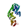
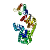
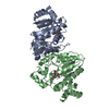
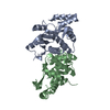
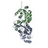
- PDB-6p5w: Structure of DCN1 bound to 3-methyl-N-((4S,5S)-3-methyl-6-oxo-1-p... -
+ Open data

- Basic information
| Entry | Database: PDB / ID: 6p5w | ||||||
|---|---|---|---|---|---|---|---|
| Title | Structure of DCN1 bound to 3-methyl-N-((4S,5S)-3-methyl-6-oxo-1-phenyl-4-(p-tolyl)-4,5,6,7-tetrahydro-1H-pyrazolo[3,4-b]pyridin-5-yl)benzamide | ||||||
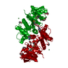
 Components Components | Lysozyme,DCN1-like protein 1 chimera | ||||||
 Keywords Keywords | LIGASE / E3 Ligase / HYDROLASE | ||||||
| Function / homology |  Function and homology information Function and homology informationpositive regulation of protein neddylation / ubiquitin-like protein binding / regulation of protein neddylation / protein neddylation / ubiquitin conjugating enzyme binding / regulation of protein ubiquitination / cullin family protein binding / ubiquitin ligase complex / viral release from host cell by cytolysis / peptidoglycan catabolic process ...positive regulation of protein neddylation / ubiquitin-like protein binding / regulation of protein neddylation / protein neddylation / ubiquitin conjugating enzyme binding / regulation of protein ubiquitination / cullin family protein binding / ubiquitin ligase complex / viral release from host cell by cytolysis / peptidoglycan catabolic process / cell wall macromolecule catabolic process / lysozyme / lysozyme activity / Neddylation / host cell cytoplasm / defense response to bacterium / nucleoplasm / nucleus / cytoplasm / cytosol Similarity search - Function | ||||||
| Biological species |  Enterobacteria phage T4 (virus) Enterobacteria phage T4 (virus) Homo sapiens (human) Homo sapiens (human) | ||||||
| Method |  X-RAY DIFFRACTION / SYNCHROTRON / MOLECULAR REPLACEMENT / Resolution: 1.69 Å X-RAY DIFFRACTION / SYNCHROTRON / MOLECULAR REPLACEMENT / Resolution: 1.69 Å | ||||||
 Authors Authors | Guy, R.K. / Kim, H.S. / Hammill, J.T. / Scott, D.C. / Schulman, B.A. | ||||||
| Funding support |  United States, 1items United States, 1items
| ||||||
 Citation Citation | Journal: J.Med.Chem. / Year: 2019 Title: Discovery of Novel Pyrazolo-pyridone DCN1 Inhibitors Controlling Cullin Neddylation. Authors: Kim, H.S. / Hammill, J.T. / Scott, D.C. / Chen, Y. / Min, J. / Rector, J. / Singh, B. / Schulman, B.A. / Guy, R.K. | ||||||
| History |
|
- Structure visualization
| Structure viewer | Molecule: MolmilJmol/JSmol |
|---|
- Downloads & links
-Download
| PDBx/mmCIF format | 6p5w.cif.gz | 231.7 KB | Display | PDBx/mmCIF format |
|---|---|---|---|---|
| PDB format | pdb6p5w.ent.gz | 185.9 KB | Display | PDB format |
| PDBx/mmJSON format | 6p5w.json.gz | Tree view | PDBx/mmJSON format | |
| Others |  Other downloads Other downloads |
-Validation report
| Arichive directory | https://data.pdbj.org/pub/pdb/validation_reports/p5/6p5wftp://data.pdbj.org/pub/pdb/validation_reports/p5/6p5w | HTTPS FTP |
|---|
-Related structure data
| Related structure data | 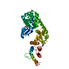 6p5vC  5v86S S: Starting model for refinement C: citing same article ( |
|---|---|
| Similar structure data |


-Links
 PDBj
PDBj


- Assembly
| Deposited unit | 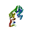
| ||||||||
|---|---|---|---|---|---|---|---|---|---|
| 1 |
| ||||||||
| Unit cell |
|
-Components
| #1: Protein | Mass: 44307.352 Da / Num. of mol.: 1 Source method: isolated from a genetically manipulated source Source: (gene. exp.) Enterobacteria phage T4 (virus), (gene. exp.) Homo sapiens (human)Gene: e, T4Tp126, DCUN1D1, DCUN1L1, RP42, SCCRO / Plasmid: pRSF DUET / Production host:  |
|---|---|
| #2: Chemical | ChemComp-O0A / 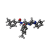  Mass: 450.532 Da / Num. of mol.: 1 / Source method: obtained synthetically / Formula: C28H26N4O2 / Feature type: SUBJECT OF INVESTIGATION Mass: 450.532 Da / Num. of mol.: 1 / Source method: obtained synthetically / Formula: C28H26N4O2 / Feature type: SUBJECT OF INVESTIGATION |
| #3: Water | ChemComp-HOH /  Mass: 18.015 Da / Num. of mol.: 327 / Source method: isolated from a natural source / Formula: H2O Mass: 18.015 Da / Num. of mol.: 327 / Source method: isolated from a natural source / Formula: H2O |
-Experimental details
-Experiment
| Experiment | Method: X-RAY DIFFRACTION / Number of used crystals: 1 |
|---|
- Sample preparation
| Crystal | Density Matthews: 2.12 Å3/Da / Density % sol: 42.04 % |
|---|---|
| Crystal grow | Temperature: 277 K / Method: vapor diffusion, hanging drop / pH: 7.5 / Details: 6% PEG3350, 0.2M NH4Br |
-Data collection
| Diffraction | Mean temperature: 100 K / Serial crystal experiment: N | |||||||||||||||||||||||||||||||||||||||||||||||||||||||||||||||||||||||||||||||||||||||||||||||||||
|---|---|---|---|---|---|---|---|---|---|---|---|---|---|---|---|---|---|---|---|---|---|---|---|---|---|---|---|---|---|---|---|---|---|---|---|---|---|---|---|---|---|---|---|---|---|---|---|---|---|---|---|---|---|---|---|---|---|---|---|---|---|---|---|---|---|---|---|---|---|---|---|---|---|---|---|---|---|---|---|---|---|---|---|---|---|---|---|---|---|---|---|---|---|---|---|---|---|---|---|---|
| Diffraction source | Source: SYNCHROTRON / Site: ALS / Beamline: 8.2.1 / Wavelength: 0.97857 Å | |||||||||||||||||||||||||||||||||||||||||||||||||||||||||||||||||||||||||||||||||||||||||||||||||||
| Detector | Type: ADSC QUANTUM 315r / Detector: CCD / Date: Oct 30, 2013 | |||||||||||||||||||||||||||||||||||||||||||||||||||||||||||||||||||||||||||||||||||||||||||||||||||
| Radiation | Protocol: SINGLE WAVELENGTH / Monochromatic (M) / Laue (L): M / Scattering type: x-ray | |||||||||||||||||||||||||||||||||||||||||||||||||||||||||||||||||||||||||||||||||||||||||||||||||||
| Radiation wavelength | Wavelength: 0.97857 Å / Relative weight: 1 | |||||||||||||||||||||||||||||||||||||||||||||||||||||||||||||||||||||||||||||||||||||||||||||||||||
| Reflection | Resolution: 1.69→50 Å / Num. obs: 40350 / % possible obs: 97.7 % / Redundancy: 3 % / Biso Wilson estimate: 23.28 Å2 / Rmerge(I) obs: 0.083 / Rpim(I) all: 0.057 / Rrim(I) all: 0.101 / Χ2: 4.211 / Net I/σ(I): 28.1 / Num. measured all: 123063 | |||||||||||||||||||||||||||||||||||||||||||||||||||||||||||||||||||||||||||||||||||||||||||||||||||
| Reflection shell | Diffraction-ID: 1
|
- Processing
| Software |
| |||||||||||||||||||||||||||||||||||||||||||||||||||||||||||||||||||||||||||||||||||||||||||||||||||||||||
|---|---|---|---|---|---|---|---|---|---|---|---|---|---|---|---|---|---|---|---|---|---|---|---|---|---|---|---|---|---|---|---|---|---|---|---|---|---|---|---|---|---|---|---|---|---|---|---|---|---|---|---|---|---|---|---|---|---|---|---|---|---|---|---|---|---|---|---|---|---|---|---|---|---|---|---|---|---|---|---|---|---|---|---|---|---|---|---|---|---|---|---|---|---|---|---|---|---|---|---|---|---|---|---|---|---|---|
| Refinement | Method to determine structure: MOLECULAR REPLACEMENT Starting model: 5V86 Resolution: 1.69→33.011 Å / SU ML: 0.16 / Cross valid method: THROUGHOUT / σ(F): 1.38 / Phase error: 20.68
| |||||||||||||||||||||||||||||||||||||||||||||||||||||||||||||||||||||||||||||||||||||||||||||||||||||||||
| Solvent computation | Shrinkage radii: 0.9 Å / VDW probe radii: 1.11 Å | |||||||||||||||||||||||||||||||||||||||||||||||||||||||||||||||||||||||||||||||||||||||||||||||||||||||||
| Displacement parameters | Biso max: 66.03 Å2 / Biso mean: 29.5509 Å2 / Biso min: 4.14 Å2 | |||||||||||||||||||||||||||||||||||||||||||||||||||||||||||||||||||||||||||||||||||||||||||||||||||||||||
| Refinement step | Cycle: final / Resolution: 1.69→33.011 Å
| |||||||||||||||||||||||||||||||||||||||||||||||||||||||||||||||||||||||||||||||||||||||||||||||||||||||||
| LS refinement shell | Refine-ID: X-RAY DIFFRACTION / Rfactor Rfree error: 0 / Total num. of bins used: 14
|
