response to methotrexate / dihydrofolate reductase / dihydrofolate reductase activity / tetrahydrofolate biosynthetic process / one-carbon metabolic process / response to xenobiotic stimulus / response to antibiotic 類似検索 - 分子機能
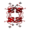
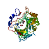
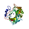
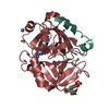
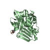
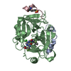
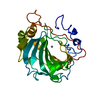
Dihydrofolate reductase, type II / R67 dihydrofolate reductase / SH3 type barrels. - #60 / Mechanosensitive ion channel MscS, beta-domain superfamily / Electron transport accessory-like domain superfamily / SH3 type barrels. / Roll / Mainly Beta 類似検索 - ドメイン・相同性
Chem-LBA / PHOSPHATE ION / Dihydrofolate reductase type 2 類似検索 - 構成要素
温度: 277 K / 手法: 蒸気拡散法, ハンギングドロップ法 / pH: 8 詳細: The protein was concentrated to 20 mg/mL in 100 mM Tris pH 8.0. Immediately before crystallization, chymotrypsin was added to the sample in a ratio of 1:100 chymotrypsin:protein, and the ...詳細: The protein was concentrated to 20 mg/mL in 100 mM Tris pH 8.0. Immediately before crystallization, chymotrypsin was added to the sample in a ratio of 1:100 chymotrypsin:protein, and the protein was diluted to 15 mg/mL using MPD, resulting in a final MPD concentration of 25%. Reservoirs were prepared using 750 uL of 100 mM sodium phosphate pH 7.6 and 60% MPD in a Greiner 24-well hanging-drop crystallization plate. On a siliconized glass cover slip (Hampton Research), 1.5 uL of protein were combined with 2.5 uL of reservoir solution and suspended over the well. The plate was incubated at 277 K and crystals were obtained in a few days.
モノクロメーター: ACCEL/BRUKER double crystal monochromator (DCM), featuring indirectly cryo-cooled first crystal and sagittally focusing second crystal プロトコル: SINGLE WAVELENGTH / 単色(M)・ラウエ(L): M / 散乱光タイプ: x-ray
解像度: 1.4→41.09 Å / Cor.coef. Fo:Fc: 0.975 / Cor.coef. Fo:Fc free: 0.975 / SU B: 0.725 / SU ML: 0.029 / SU R Cruickshank DPI: 0.0493 / 交差検証法: THROUGHOUT / σ(F): 0 / ESU R: 0.049 / ESU R Free: 0.049 詳細: Authors state that inhibitor LBA is partially modelled at the active site. Binding of the ligand has been confirmed biochemically, and ligand disorder outside of the pore center has been ...詳細: Authors state that inhibitor LBA is partially modelled at the active site. Binding of the ligand has been confirmed biochemically, and ligand disorder outside of the pore center has been observed for other known ligands. More details can be found in the primary citation.
 ムービー
ムービー コントローラー


 データを開く
 基本情報
基本情報 要素
要素 キーワード
キーワード 機能・相同性情報
 X線回折 /
  データ登録者
 カナダ, 5件
  引用
引用 構造の表示
構造の表示 ダウンロードとリンク
 その他のダウンロード
 PDBj
PDBj


 集合体
集合体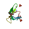

 分子量: 564.545 Da / 分子数: 1 / 由来タイプ: 合成 / 式: C31H24N4O7 / タイプ: SUBJECT OF INVESTIGATION
 分子量: 118.174 Da / 分子数: 2 / 由来タイプ: 合成 / 式: C6H14O2 / コメント: 沈殿剤*YM
 分子量: 94.971 Da / 分子数: 2 / 由来タイプ: 合成 / 式: PO4
 分子量: 18.015 Da / 分子数: 64 / 由来タイプ: 天然 / 式: H2O
 試料調製
試料調製 解析
解析