 Movie
Movie Controller

[English] 日本語
 Yorodumi
- PDB-6huz: HmdII from Desulfurobacterium thermolithotrophum reconstituted wi... -
+ Open data

- Basic information
| Entry | Database: PDB / ID: 6huz | |||||||||
|---|---|---|---|---|---|---|---|---|---|---|
| Title | HmdII from Desulfurobacterium thermolithotrophum reconstituted with Fe-guanylylpyridinol (FeGP) cofactor and co-crystallized with methenyl-tetrahydrofolate form B | |||||||||
 Components Components | Coenzyme F420-dependent N(5),N(10)-methenyltetrahydromethanopterin reductase-related protein | |||||||||
 Keywords Keywords | OXIDOREDUCTASE / Hydrogenase / H2-activation / Lateral gene-transfer / cofactor biosynthesis / tetrahydromethanopterin / tetrahydrofolate / paralog / sulfur-reducing bacteria / metalloenzyme | |||||||||
| Function / homology | H(2)-forming methylenetetrahydromethanopterin dehydrogenase-related protein / H2-forming N5,N10-methylenetetrahydromethanopterin dehydrogenase, C-terminal / HMD, C-terminal domain superfamily / H2-forming N5,N10-methylene-tetrahydromethanopterin dehydrogenase / 6-phosphogluconate dehydrogenase-like, C-terminal domain superfamily / NAD(P)-binding domain superfamily / iron-guanylyl pyridinol cofactor / 5,10-Methenyltetrahydrofolate / Coenzyme F420-dependent N(5),N(10)-methenyltetrahydromethanopterin reductase-related protein Function and homology information Function and homology information | |||||||||
| Biological species |  Desulfurobacterium thermolithotrophum DSM 11699 (bacteria) Desulfurobacterium thermolithotrophum DSM 11699 (bacteria) | |||||||||
| Method |  X-RAY DIFFRACTION / SYNCHROTRON / MOLECULAR REPLACEMENT / Resolution: 1.85 Å X-RAY DIFFRACTION / SYNCHROTRON / MOLECULAR REPLACEMENT / Resolution: 1.85 Å | |||||||||
 Authors Authors | Watanabe, T. / Wagner, T. / Huang, G. / Kahnt, J. / Ataka, K. / Ermler, U. / Shima, S. | |||||||||
| Funding support |  Germany, Germany,  China, 2items China, 2items
| |||||||||
 Citation Citation | Journal: Angew. Chem. Int. Ed. Engl. / Year: 2019 Title: The Bacterial [Fe]-Hydrogenase Paralog HmdII Uses Tetrahydrofolate Derivatives as Substrates. Authors: Watanabe, T. / Wagner, T. / Huang, G. / Kahnt, J. / Ataka, K. / Ermler, U. / Shima, S. | |||||||||
| History |
|
- Structure visualization
| Structure viewer | Molecule: MolmilJmol/JSmol |
|---|
- Downloads & links
-Download
| PDBx/mmCIF format | 6huz.cif.gz | 165.6 KB | Display | PDBx/mmCIF format |
|---|---|---|---|---|
| PDB format | pdb6huz.ent.gz | 127.9 KB | Display | PDB format |
| PDBx/mmJSON format | 6huz.json.gz | Tree view | PDBx/mmJSON format | |
| Others |  Other downloads Other downloads |
-Validation report
| Summary document | 6huz_validation.pdf.gz | 985.8 KB | Display | wwPDB validaton report |
|---|---|---|---|---|
| Full document | 6huz_full_validation.pdf.gz | 987.3 KB | Display | |
| Data in XML | 6huz_validation.xml.gz | 17.8 KB | Display | |
| Data in CIF | 6huz_validation.cif.gz | 26.6 KB | Display | |
| Arichive directory | https://data.pdbj.org/pub/pdb/validation_reports/hu/6huzftp://data.pdbj.org/pub/pdb/validation_reports/hu/6huz | HTTPS FTP |
-Related structure data
| Related structure data |  6huxC  6huySC S: Starting model for refinement C: citing same article ( |
|---|---|
| Similar structure data |


-Links
 PDBj
PDBj


- Assembly
| Deposited unit | 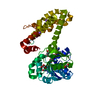
| ||||||||
|---|---|---|---|---|---|---|---|---|---|
| 1 | 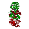
| ||||||||
| Unit cell |
|
-Components
-Protein , 1 types, 1 molecules A
| #1: Protein | Mass: 40994.000 Da / Num. of mol.: 1 / Mutation: wild-type Source method: isolated from a genetically manipulated source Details: synthetic gene Source: (gene. exp.) Desulfurobacterium thermolithotrophum DSM 11699 (bacteria)Tissue: / / Cell: / / Cell line: / / Gene: Dester_1504 / Organ: / / Details (production host): / / Cell (production host): / / Organ (production host): / / Production host: |
|---|
-Non-polymers , 6 types, 289 molecules 


| #2: Chemical | ChemComp-FE9 /  Mass: 686.323 Da / Num. of mol.: 1 / Source method: obtained synthetically / Formula: C21H23FeN6O13PS Mass: 686.323 Da / Num. of mol.: 1 / Source method: obtained synthetically / Formula: C21H23FeN6O13PS | ||||||
|---|---|---|---|---|---|---|---|
| #3: Chemical | ChemComp-GUE /  Mass: 456.432 Da / Num. of mol.: 1 / Source method: obtained synthetically / Formula: C20H22N7O6 Mass: 456.432 Da / Num. of mol.: 1 / Source method: obtained synthetically / Formula: C20H22N7O6 | ||||||
| #4: Chemical |  Mass: 92.094 Da / Num. of mol.: 2 / Source method: obtained synthetically / Formula: C3H8O3 Mass: 92.094 Da / Num. of mol.: 2 / Source method: obtained synthetically / Formula: C3H8O3#5: Chemical | ChemComp-EDO / |  Mass: 62.068 Da / Num. of mol.: 1 / Source method: obtained synthetically / Formula: C2H6O2 Mass: 62.068 Da / Num. of mol.: 1 / Source method: obtained synthetically / Formula: C2H6O2#6: Chemical |  Mass: 22.990 Da / Num. of mol.: 3 / Source method: obtained synthetically / Formula: Na Mass: 22.990 Da / Num. of mol.: 3 / Source method: obtained synthetically / Formula: Na#7: Water | ChemComp-HOH / | Mass: 18.015 Da / Num. of mol.: 281 / Source method: isolated from a natural source / Formula: H2O |
-Experimental details
-Experiment
| Experiment | Method: X-RAY DIFFRACTION / Number of used crystals: 1 |
|---|
- Sample preparation
| Crystal | Density Matthews: 2.58 Å3/Da / Density % sol: 52.43 % / Description: Transparent triangular shape |
|---|---|
| Crystal grow | Temperature: 281 K / Method: vapor diffusion, sitting drop / pH: 5.5 Details: HmdII from Desulfurobacterium thermolithotrophum was reconstituted with the Fe-guanylylpyridinol cofactor from Methanothermobacter marburgensis and co-crystallized with methenyl- ...Details: HmdII from Desulfurobacterium thermolithotrophum was reconstituted with the Fe-guanylylpyridinol cofactor from Methanothermobacter marburgensis and co-crystallized with methenyl-tetrahydrofolate using the sitting drop vapor diffusion method under N2/H2 (95%/5%) in red light condition. The reconstituted holoenzyme was mixed with methenyl-H4F+ at the final concentrations of 1.6 mM. Methenyl-tetrahydrofolate was dissolved in 10% DMSO, and the final concentration of DMSO in the protein solution was 1.6%. 0.7 ul of dHmdII at 21 mg/ml (reconstituted with FeGP and methenyl-H4F+) was spotted on a 96-well 2-drop MRC Crystallization Plates (Molecular Dimensions, Suffolk, UK) and 0.7 ul of reservoir solution was mixed. After one month, crystals appeared in 20% PEG 3000 (w/v) and 100 mM Sodium Citrate pH 5.5 (from the kit WIZARD, Jena Bioscience). The crystals were cryoprotected by a soak of few seconds in 20% PEG 3000 (w/v), 100 mM Tri-Sodium Citrate pH 5.5 and 20% glycerol before a flash freeze in liquid nitrogen Temp details: The temperature was fluctuating by +/- 1 degree |
-Data collection
| Diffraction | Mean temperature: 100 K / Serial crystal experiment: N |
|---|---|
| Diffraction source | Source: SYNCHROTRON / Site: SOLEIL  / Beamline: PROXIMA 1 / Wavelength: 0.97857 Å / Beamline: PROXIMA 1 / Wavelength: 0.97857 Å |
| Detector | Type: DECTRIS PILATUS 6M / Detector: PIXEL / Date: Jun 7, 2018 |
| Radiation | Protocol: SINGLE WAVELENGTH / Monochromatic (M) / Laue (L): M / Scattering type: x-ray |
| Radiation wavelength | Wavelength: 0.97857 Å / Relative weight: 1 |
| Reflection | Resolution: 1.85→49.16 Å / Num. obs: 37060 / % possible obs: 99.9 % / Redundancy: 6.5 % / Biso Wilson estimate: 32.15 Å2 / CC1/2: 0.998 / Rmerge(I) obs: 0.085 / Rpim(I) all: 0.036 / Rrim(I) all: 0.092 / Net I/σ(I): 10.2 |
| Reflection shell | Resolution: 1.85→1.95 Å / Redundancy: 6.6 % / Rmerge(I) obs: 1.212 / Mean I/σ(I) obs: 1.2 / Num. unique obs: 5308 / CC1/2: 0.849 / Rpim(I) all: 0.511 / Rrim(I) all: 1.317 / % possible all: 99.7 |
- Processing
| Software |
| ||||||||||||||||||||||||||||||||||||||||||||||||||||||||||||||||||||||||||||||||||||||||||||||||||||||||||||||||||
|---|---|---|---|---|---|---|---|---|---|---|---|---|---|---|---|---|---|---|---|---|---|---|---|---|---|---|---|---|---|---|---|---|---|---|---|---|---|---|---|---|---|---|---|---|---|---|---|---|---|---|---|---|---|---|---|---|---|---|---|---|---|---|---|---|---|---|---|---|---|---|---|---|---|---|---|---|---|---|---|---|---|---|---|---|---|---|---|---|---|---|---|---|---|---|---|---|---|---|---|---|---|---|---|---|---|---|---|---|---|---|---|---|---|---|---|
| Refinement | Method to determine structure: MOLECULAR REPLACEMENT Starting model: 6HUY Resolution: 1.85→46.39 Å / Cor.coef. Fo:Fc: 0.959 / Cor.coef. Fo:Fc free: 0.951 / SU R Cruickshank DPI: 0.182 / Cross valid method: THROUGHOUT / σ(F): 0 / SU R Blow DPI: 0.139 / SU Rfree Blow DPI: 0.125 / SU Rfree Cruickshank DPI: 0.121 Details: The C-terminal helix 335-347 might be in two different position and only the one with the highest occupancy has been modelled. The final part 350-356 has been traced to fit an extra electron ...Details: The C-terminal helix 335-347 might be in two different position and only the one with the highest occupancy has been modelled. The final part 350-356 has been traced to fit an extra electron density, however it is not excluded that this density corresponds to polyethylene glycol. The hydrogens have been added in riding position for the last refinement cycle and have been omitted in the deposited model.
| ||||||||||||||||||||||||||||||||||||||||||||||||||||||||||||||||||||||||||||||||||||||||||||||||||||||||||||||||||
| Displacement parameters | Biso mean: 44.11 Å2
| ||||||||||||||||||||||||||||||||||||||||||||||||||||||||||||||||||||||||||||||||||||||||||||||||||||||||||||||||||
| Refine analyze | Luzzati coordinate error obs: 0.25 Å | ||||||||||||||||||||||||||||||||||||||||||||||||||||||||||||||||||||||||||||||||||||||||||||||||||||||||||||||||||
| Refinement step | Cycle: 1 / Resolution: 1.85→46.39 Å
| ||||||||||||||||||||||||||||||||||||||||||||||||||||||||||||||||||||||||||||||||||||||||||||||||||||||||||||||||||
| Refine LS restraints |
| ||||||||||||||||||||||||||||||||||||||||||||||||||||||||||||||||||||||||||||||||||||||||||||||||||||||||||||||||||
| LS refinement shell | Resolution: 1.85→1.9 Å / Total num. of bins used: 18
| ||||||||||||||||||||||||||||||||||||||||||||||||||||||||||||||||||||||||||||||||||||||||||||||||||||||||||||||||||
| Refinement TLS params. | Method: refined / Origin x: -31.3793 Å / Origin y: 15.6653 Å / Origin z: 14.6374 Å
| ||||||||||||||||||||||||||||||||||||||||||||||||||||||||||||||||||||||||||||||||||||||||||||||||||||||||||||||||||
| Refinement TLS group | Selection details: { A|* } |
