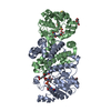
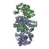
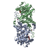
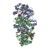
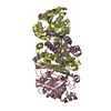
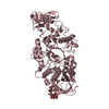
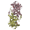
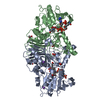
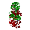
- PDB-6huz: HmdII from Desulfurobacterium thermolithotrophum reconstituted wi... -
+
データを開く
IDまたはキーワード:
読み込み中...
-
基本情報
登録情報
データベース: PDB / ID: 6huz
タイトル
HmdII from Desulfurobacterium thermolithotrophum reconstituted with Fe-guanylylpyridinol (FeGP) cofactor and co-crystallized with methenyl-tetrahydrofolate form B
要素
Coenzyme F420-dependent N(5),N(10)-methenyltetrahydromethanopterin reductase-related protein
温度: 281 K / 手法: 蒸気拡散法, シッティングドロップ法 / pH: 5.5 詳細: HmdII from Desulfurobacterium thermolithotrophum was reconstituted with the Fe-guanylylpyridinol cofactor from Methanothermobacter marburgensis and co-crystallized with methenyl- ...詳細: HmdII from Desulfurobacterium thermolithotrophum was reconstituted with the Fe-guanylylpyridinol cofactor from Methanothermobacter marburgensis and co-crystallized with methenyl-tetrahydrofolate using the sitting drop vapor diffusion method under N2/H2 (95%/5%) in red light condition. The reconstituted holoenzyme was mixed with methenyl-H4F+ at the final concentrations of 1.6 mM. Methenyl-tetrahydrofolate was dissolved in 10% DMSO, and the final concentration of DMSO in the protein solution was 1.6%. 0.7 ul of dHmdII at 21 mg/ml (reconstituted with FeGP and methenyl-H4F+) was spotted on a 96-well 2-drop MRC Crystallization Plates (Molecular Dimensions, Suffolk, UK) and 0.7 ul of reservoir solution was mixed. After one month, crystals appeared in 20% PEG 3000 (w/v) and 100 mM Sodium Citrate pH 5.5 (from the kit WIZARD, Jena Bioscience). The crystals were cryoprotected by a soak of few seconds in 20% PEG 3000 (w/v), 100 mM Tri-Sodium Citrate pH 5.5 and 20% glycerol before a flash freeze in liquid nitrogen Temp details: The temperature was fluctuating by +/- 1 degree
解像度: 1.85→46.39 Å / Cor.coef. Fo:Fc: 0.959 / Cor.coef. Fo:Fc free: 0.951 / SU R Cruickshank DPI: 0.182 / 交差検証法: THROUGHOUT / σ(F): 0 / SU R Blow DPI: 0.139 / SU Rfree Blow DPI: 0.125 / SU Rfree Cruickshank DPI: 0.121 詳細: The C-terminal helix 335-347 might be in two different position and only the one with the highest occupancy has been modelled. The final part 350-356 has been traced to fit an extra electron ...詳細: The C-terminal helix 335-347 might be in two different position and only the one with the highest occupancy has been modelled. The final part 350-356 has been traced to fit an extra electron density, however it is not excluded that this density corresponds to polyethylene glycol. The hydrogens have been added in riding position for the last refinement cycle and have been omitted in the deposited model.
Rfactor
反射数
%反射
Selection details
Rfree
0.223
1812
4.97 %
RANDOM
Rwork
0.195
-
-
-
obs
0.196
36480
98.4 %
-
原子変位パラメータ
Biso mean: 44.11 Å2
Baniso -1
Baniso -2
Baniso -3
1-
-1.3291 Å2
0 Å2
0 Å2
2-
-
-0.4571 Å2
0 Å2
3-
-
-
1.7863 Å2
Refine analyze
Luzzati coordinate error obs: 0.25 Å
精密化ステップ
サイクル: 1 / 解像度: 1.85→46.39 Å
タンパク質
核酸
リガンド
溶媒
全体
原子数
2739
0
93
281
3113
拘束条件
Refine-ID
タイプ
Dev ideal
数
Restraint function
Weight
X-RAY DIFFRACTION
t_bond_d
0.01
5791
HARMONIC
5
X-RAY DIFFRACTION
t_angle_deg
1.14
10552
HARMONIC
5
X-RAY DIFFRACTION
t_dihedral_angle_d
1331
SINUSOIDAL
2
X-RAY DIFFRACTION
t_incorr_chiral_ct
X-RAY DIFFRACTION
t_pseud_angle
X-RAY DIFFRACTION
t_trig_c_planes
X-RAY DIFFRACTION
t_gen_planes
864
HARMONIC
30
X-RAY DIFFRACTION
t_it
5791
HARMONIC
20
X-RAY DIFFRACTION
t_nbd
3
SEMIHARMONIC
5
X-RAY DIFFRACTION
t_omega_torsion
1.75
X-RAY DIFFRACTION
t_other_torsion
15.23
X-RAY DIFFRACTION
t_improper_torsion
X-RAY DIFFRACTION
t_chiral_improper_torsion
396
SEMIHARMONIC
5
X-RAY DIFFRACTION
t_sum_occupancies
X-RAY DIFFRACTION
t_utility_distance
X-RAY DIFFRACTION
t_utility_angle
X-RAY DIFFRACTION
t_utility_torsion
X-RAY DIFFRACTION
t_ideal_dist_contact
6579
SEMIHARMONIC
4
LS精密化 シェル
解像度: 1.85→1.9 Å / Total num. of bins used: 18
Rfactor
反射数
%反射
Rfree
0.3479
145
5.07 %
Rwork
0.3009
2717
-
all
0.3032
2862
-
obs
-
-
95.39 %
精密化 TLS
手法: refined / Origin x: -31.3793 Å / Origin y: 15.6653 Å / Origin z: 14.6374 Å
 ムービー
ムービー コントローラー


 データを開く
 基本情報
基本情報 要素
要素 キーワード
キーワード 機能・相同性情報
 Desulfurobacterium thermolithotrophum DSM 11699 (バクテリア)
 X線回折 /
  データ登録者
 ドイツ,
ドイツ,  中国, 2件
  引用
引用 構造の表示
構造の表示 ダウンロードとリンク
 その他のダウンロード


 PDBj
PDBj


 集合体
集合体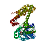
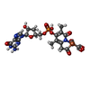
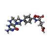


 分子量: 686.323 Da / 分子数: 1 / 由来タイプ: 合成 / 式: C21H23FeN6O13PS
 分子量: 456.432 Da / 分子数: 1 / 由来タイプ: 合成 / 式: C20H22N7O6
 分子量: 92.094 Da / 分子数: 2 / 由来タイプ: 合成 / 式: C3H8O3
 分子量: 62.068 Da / 分子数: 1 / 由来タイプ: 合成 / 式: C2H6O2
 分子量: 22.990 Da / 分子数: 3 / 由来タイプ: 合成 / 式: Na
 試料調製
試料調製 / ビームライン: PROXIMA 1 / 波長: 0.97857 Å
 解析
解析