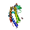
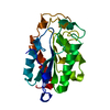
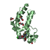
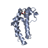
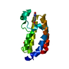
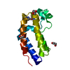
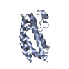
Entry Database : PDB / ID : 6fffTitle Human BRD2 C-terminal bromodomain with (S)-5-(1-acetyl-2-cyclopropyl-4-(2-(hydroxymethyl)benzyl)-1,2,3,4-tetrahydroquinoxalin-6-yl)pyrimidine-2-carboxamide Bromodomain-containing protein 2 Keywords / / / / / / / Function / homology Function Domain/homology Component
/ / / / / / / / / / / / / / / / / / / / / / / / / / / / / / / / / / / / / / / / / / / / / / / / / / / / / / / Biological species Homo sapiens (human)Method / / Resolution : 1.67 Å Authors Chung, C. Journal : J.Med.Chem. / Year : 2018Title : Discovery of Tetrahydroquinoxalines as Bromodomain and Extra-Terminal Domain (BET) Inhibitors with Selectivity for the Second Bromodomain.Authors : Law, R.P. / Atkinson, S.J. / Bamborough, P. / Chung, C.W. / Demont, E.H. / Gordon, L.J. / Lindon, M. / Prinjha, R.K. / Watson, A.J.B. / Hirst, D.J. History Deposition Jan 7, 2018 Deposition site / Processing site Revision 1.0 Jan 30, 2019 Provider / Type Revision 1.1 Feb 12, 2020 Group / Category / citation_authorItem _citation.country / _citation.journal_abbrev ... _citation.country / _citation.journal_abbrev / _citation.journal_id_ASTM / _citation.journal_id_CSD / _citation.journal_id_ISSN / _citation.journal_volume / _citation.page_first / _citation.page_last / _citation.pdbx_database_id_DOI / _citation.pdbx_database_id_PubMed / _citation.title / _citation.year Revision 1.2 May 8, 2024 Group / Database references / Category / chem_comp_bond / database_2Item / _database_2.pdbx_database_accession
Show all Show less
 Movie
Movie Controller

 Yorodumi
 Open data
 Basic information
 Components
 Keywords
 Function and homology information
 Homo sapiens (human)
 X-RAY DIFFRACTION /
  Authors
 Citation
 Structure visualization
 Downloads & links
 Other downloads
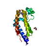


 PDBj
PDBj

 Assembly
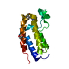


 Mass: 62.068 Da / Num. of mol.: 1 / Source method: obtained synthetically / Formula: C2H6O2
 Mass: 195.237 Da / Num. of mol.: 1 / Source method: obtained synthetically / Formula: C6H13NO4S / Comment: pH buffer*YM
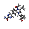
 Mass: 457.524 Da / Num. of mol.: 1 / Source method: obtained synthetically / Formula: C26H27N5O3
 Mass: 18.015 Da / Num. of mol.: 278 / Source method: isolated from a natural source / Formula: H2O
 Sample preparation
 Processing