 Movie
Movie Controller


+ Open data

- Basic information
| Entry | Database: PDB / ID: 6et8 | ||||||
|---|---|---|---|---|---|---|---|
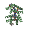
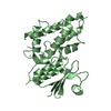
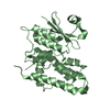
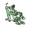
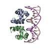
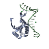
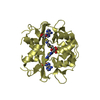
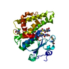
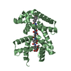
| Title | Crystal structure of AlbA in complex with albicidin | ||||||
 Components Components | Albicidin resistance protein | ||||||
 Keywords Keywords | PROTEIN BINDING / albicidin / AlbA | ||||||
| Function / homology | TipAS antibiotic-recognition domain / TipAS antibiotic-recognition domain / albicidin / Albicidin resistance protein Function and homology information Function and homology information | ||||||
| Biological species |  Klebsiella oxytoca (bacteria) Klebsiella oxytoca (bacteria) | ||||||
| Method |  X-RAY DIFFRACTION / SYNCHROTRON / SAD / Resolution: 1.7 Å X-RAY DIFFRACTION / SYNCHROTRON / SAD / Resolution: 1.7 Å | ||||||
 Authors Authors | Driller, R. / Rostock, L. / Alings, C. / Graetz, S. / Suessmuth, R. / Mainz, A. / Wahl, M.C. / Loll, B. | ||||||
 Citation Citation | Journal: Nat Commun / Year: 2018 Title: Molecular insights into antibiotic resistance - how a binding protein traps albicidin. Authors: Rostock, L. / Driller, R. / Gratz, S. / Kerwat, D. / von Eckardstein, L. / Petras, D. / Kunert, M. / Alings, C. / Schmitt, F.J. / Friedrich, T. / Wahl, M.C. / Loll, B. / Mainz, A. / Sussmuth, R.D. | ||||||
| History |
|
- Structure visualization
| Structure viewer | Molecule: MolmilJmol/JSmol |
|---|
- Downloads & links
-Download
| PDBx/mmCIF format | 6et8.cif.gz | 207.1 KB | Display | PDBx/mmCIF format |
|---|---|---|---|---|
| PDB format | pdb6et8.ent.gz | 176.7 KB | Display | PDB format |
| PDBx/mmJSON format | 6et8.json.gz | Tree view | PDBx/mmJSON format | |
| Others |  Other downloads Other downloads |
-Validation report
| Summary document | 6et8_validation.pdf.gz | 815.6 KB | Display | wwPDB validaton report |
|---|---|---|---|---|
| Full document | 6et8_full_validation.pdf.gz | 819.9 KB | Display | |
| Data in XML | 6et8_validation.xml.gz | 22.4 KB | Display | |
| Data in CIF | 6et8_validation.cif.gz | 32.7 KB | Display | |
| Arichive directory | https://data.pdbj.org/pub/pdb/validation_reports/et/6et8ftp://data.pdbj.org/pub/pdb/validation_reports/et/6et8 | HTTPS FTP |
-Related structure data
| Similar structure data | |
|---|---|
| Experimental dataset #1 | Data reference: 10.18430/m36et8 / Data set type: diffraction image data |

-Links
 PDBj
PDBj- Assembly
| Deposited unit | 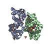
| |||||||||
|---|---|---|---|---|---|---|---|---|---|---|
| 1 | 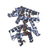
| |||||||||
| 2 | 
| |||||||||
| Unit cell |
| |||||||||
| Components on special symmetry positions |
|
-Components
| #1: Protein | Mass: 26581.107 Da / Num. of mol.: 2 Source method: isolated from a genetically manipulated source Details: First residues is an artefact of the tag sequence. SeMet-labelled protein has been crystallised. Source: (gene. exp.) Klebsiella oxytoca (bacteria) / Gene: albA / Production host: #2: Chemical |   Mass: 842.806 Da / Num. of mol.: 2 / Source method: obtained synthetically / Formula: C44H38N6O12 Mass: 842.806 Da / Num. of mol.: 2 / Source method: obtained synthetically / Formula: C44H38N6O12#3: Chemical | ChemComp-SO4 / |   Mass: 96.063 Da / Num. of mol.: 1 / Source method: obtained synthetically / Formula: SO4 Mass: 96.063 Da / Num. of mol.: 1 / Source method: obtained synthetically / Formula: SO4#4: Water | ChemComp-HOH / |  Mass: 18.015 Da / Num. of mol.: 372 / Source method: isolated from a natural source / Formula: H2O Mass: 18.015 Da / Num. of mol.: 372 / Source method: isolated from a natural source / Formula: H2O |
|---|
-Experimental details
-Experiment
| Experiment | Method: X-RAY DIFFRACTION / Number of used crystals: 1 |
|---|
- Sample preparation
| Crystal | Density Matthews: 2.56 Å3/Da / Density % sol: 51.9 % |
|---|---|
| Crystal grow | Temperature: 291.15 K / Method: vapor diffusion, sitting drop / Details: 0.1 M Tris pH 8.5 2.0 M ammonium sulfate |
-Data collection
| Diffraction | Mean temperature: 100 K |
|---|---|
| Diffraction source | Source: SYNCHROTRON / Site: BESSY  / Beamline: 14.1 / Wavelength: 0.97949 Å / Beamline: 14.1 / Wavelength: 0.97949 Å |
| Detector | Type: DECTRIS PILATUS 6M / Detector: PIXEL / Date: Mar 9, 2017 |
| Radiation | Monochromator: SI111-DCM / Protocol: SINGLE WAVELENGTH / Monochromatic (M) / Laue (L): M / Scattering type: x-ray |
| Radiation wavelength | Wavelength: 0.97949 Å / Relative weight: 1 |
| Reflection | Resolution: 1.7→50 Å / Num. obs: 113199 / % possible obs: 100 % / Redundancy: 13.5 % / Rrim(I) all: 0.11 / Net I/σ(I): 16.53 |
| Reflection shell | Resolution: 1.7→1.74 Å / Redundancy: 13.62 % / Mean I/σ(I) obs: 1.49 / Num. unique all: 8420 / CC1/2: 0.669 / Rrim(I) all: 1.796 / % possible all: 100 |
- Processing
| Software |
| |||||||||||||||||||||||||||||||||||||||||||||||||||||||||||||||||||||||||||||||||||||||||||||||||||||||||||||||||||||||||||||
|---|---|---|---|---|---|---|---|---|---|---|---|---|---|---|---|---|---|---|---|---|---|---|---|---|---|---|---|---|---|---|---|---|---|---|---|---|---|---|---|---|---|---|---|---|---|---|---|---|---|---|---|---|---|---|---|---|---|---|---|---|---|---|---|---|---|---|---|---|---|---|---|---|---|---|---|---|---|---|---|---|---|---|---|---|---|---|---|---|---|---|---|---|---|---|---|---|---|---|---|---|---|---|---|---|---|---|---|---|---|---|---|---|---|---|---|---|---|---|---|---|---|---|---|---|---|---|
| Refinement | Method to determine structure: SAD / Resolution: 1.7→42.048 Å / SU ML: 0.18 / Cross valid method: FREE R-VALUE / σ(F): 1.35 / Phase error: 19.59
| |||||||||||||||||||||||||||||||||||||||||||||||||||||||||||||||||||||||||||||||||||||||||||||||||||||||||||||||||||||||||||||
| Solvent computation | Shrinkage radii: 0.9 Å / VDW probe radii: 1.11 Å | |||||||||||||||||||||||||||||||||||||||||||||||||||||||||||||||||||||||||||||||||||||||||||||||||||||||||||||||||||||||||||||
| Refinement step | Cycle: LAST / Resolution: 1.7→42.048 Å
| |||||||||||||||||||||||||||||||||||||||||||||||||||||||||||||||||||||||||||||||||||||||||||||||||||||||||||||||||||||||||||||
| Refine LS restraints |
| |||||||||||||||||||||||||||||||||||||||||||||||||||||||||||||||||||||||||||||||||||||||||||||||||||||||||||||||||||||||||||||
| LS refinement shell |
| |||||||||||||||||||||||||||||||||||||||||||||||||||||||||||||||||||||||||||||||||||||||||||||||||||||||||||||||||||||||||||||
| Refinement TLS params. | Method: refined / Refine-ID: X-RAY DIFFRACTION
| |||||||||||||||||||||||||||||||||||||||||||||||||||||||||||||||||||||||||||||||||||||||||||||||||||||||||||||||||||||||||||||
| Refinement TLS group |
|
