[English] 日本語
Yorodumi
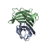
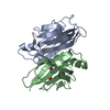
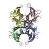
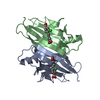
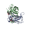
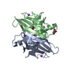
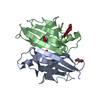
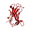
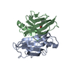
- PDB-6d0w: Structure of human transthyretin complex with analgesic inhibitor -

+
Open data


ID or keywords:

Loading...

-
Basic information

Entry
Database: PDB / ID: 6d0w
TitleStructure of human transthyretin complex with analgesic inhibitor
ComponentsTransthyretin
KeywordsProtein Transport/Protein Transport Inhibitor / transthyretin transport protein inhibitor complex / PROTEIN TRANSPORT / Protein Transport-Protein Transport Inhibitor complex
Function / homology
Function and homology information


Retinoid cycle disease events / The canonical retinoid cycle in rods (twilight vision) / thyroid hormone binding / purine nucleobase metabolic process / Non-integrin membrane-ECM interactions / Retinoid metabolism and transport / hormone activity / azurophil granule lumen / Amyloid fiber formation / Neutrophil degranulation ...Retinoid cycle disease events / The canonical retinoid cycle in rods (twilight vision) / thyroid hormone binding / purine nucleobase metabolic process / Non-integrin membrane-ECM interactions / Retinoid metabolism and transport / hormone activity / azurophil granule lumen / Amyloid fiber formation / Neutrophil degranulation / extracellular space / extracellular exosome / extracellular region / identical protein binding
Similarity search - Function
Transthyretin, conserved site / Transthyretin signature 2. / Transthyretin, thyroxine binding site / Transthyretin signature 1. / Transthyretin / Transthyretin/hydroxyisourate hydrolase / Transthyretin/hydroxyisourate hydrolase domain / Transthyretin/hydroxyisourate hydrolase domain superfamily / HIUase/Transthyretin family
Similarity search - Domain/homology
2-[(3,5-dichlorophenyl)amino]benzoic acid / Transthyretin
Similarity search - Component
Biological speciesHomo sapiens (human)
MethodX-RAY DIFFRACTION / SYNCHROTRON / MOLECULAR REPLACEMENT / Resolution: 1.7 Å
AuthorsCody, V.
CitationJournal: To Be Published
Title: Structural Analysis of Human Transthyretin Analgesic Inhibitor Complex
Authors: Cody, V. / Richardson, S.
History
DepositionApr 11, 2018Deposition site: RCSB / Processing site: RCSB
Revision 1.0Apr 17, 2019Provider: repository / Type: Initial release
Revision 1.1Oct 4, 2023Group: Data collection / Database references ...Data collection / Database references / Derived calculations / Refinement description
Category: chem_comp_atom / chem_comp_bond ...chem_comp_atom / chem_comp_bond / database_2 / pdbx_initial_refinement_model / struct_conn
Item: _database_2.pdbx_DOI / _database_2.pdbx_database_accession ..._database_2.pdbx_DOI / _database_2.pdbx_database_accession / _struct_conn.ptnr1_auth_asym_id / _struct_conn.ptnr1_auth_comp_id / _struct_conn.ptnr1_auth_seq_id / _struct_conn.ptnr1_label_asym_id / _struct_conn.ptnr1_label_atom_id / _struct_conn.ptnr1_label_comp_id / _struct_conn.ptnr1_label_seq_id / _struct_conn.ptnr2_auth_asym_id / _struct_conn.ptnr2_auth_comp_id / _struct_conn.ptnr2_auth_seq_id / _struct_conn.ptnr2_label_asym_id / _struct_conn.ptnr2_label_atom_id / _struct_conn.ptnr2_label_comp_id / _struct_conn.ptnr2_label_seq_id

-
Structure visualization

Structure viewerMolecule:
MolmilJmol/JSmol

Downloads & links

-
Assembly

Deposited unit
A: Transthyretin
B: Transthyretin
hetero molecules


Theoretical massNumber of molelcules
Total (without water)28,1405
Polymers27,5532
Non-polymers5873
Water1,874104
1
A: Transthyretin
B: Transthyretin
hetero molecules

A: Transthyretin
B: Transthyretin
hetero molecules


Theoretical massNumber of molelcules
Total (without water)56,28010
Polymers55,1064
Non-polymers1,1746
Water724
TypeNameSymmetry operationNumber
identity operation1_555x,y,z1
crystal symmetry operation2_555-x,-y,z1
Buried area8590 Å2
ΔGint-41 kcal/mol
Surface area18640 Å2
MethodPISA
Unit cell
Length a, b, c (Å)43.979, 85.405, 65.006
Angle α, β, γ (deg.)90.00, 90.00, 90.00
Int Tables number18
Space group name H-MP21212
Components on special symmetry positions
IDModelComponents
11A-201-

FQ7

21A-201-

FQ7

31A-201-

FQ7

41A-201-

FQ7

51A-201-

FQ7

61B-201-

FQ7

71B-201-

FQ7

-
Components

#1: Protein Transthyretin / ATTR / Prealbumin / TBPA


Mass: 13776.376 Da / Num. of mol.: 2 / Source method: isolated from a natural source / Source: (natural) Homo sapiens (human) / Plasmid details: human sera / References: UniProt: P02766
#2: Chemical ChemComp-FQ7 / 2-[(3,5-dichlorophenyl)amino]benzoic acid


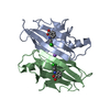
Mass: 282.122 Da / Num. of mol.: 2 / Source method: obtained synthetically / Formula: C13H9Cl2NO2
#3: Chemical ChemComp-NA / SODIUM ION


Mass: 22.990 Da / Num. of mol.: 1 / Source method: obtained synthetically / Formula: Na
#4: Water ChemComp-HOH / water


Mass: 18.015 Da / Num. of mol.: 104 / Source method: isolated from a natural source / Formula: H2O

-
Experimental details

-
Experiment

ExperimentMethod: X-RAY DIFFRACTION / Number of used crystals: 1

-
Sample preparation

CrystalDensity Matthews: 2.41 Å3/Da / Density % sol: 44.48 %
Crystal growTemperature: 293 K / Method: vapor diffusion, hanging drop / pH: 5.5 / Details: 0.1 M MES, pH 5.5, PEG 4K, 150 mM AmSO4

-
Data collection

DiffractionMean temperature: 113 K
Diffraction sourceSource: SYNCHROTRON / Site: SSRL / Beamline: BL11-1 / Wavelength: 0.987 Å
DetectorType: DECTRIS PILATUS 300K / Detector: PIXEL / Date: Jun 3, 2013
RadiationMonochromator: Zr filter / Protocol: SINGLE WAVELENGTH / Monochromatic (M) / Laue (L): M / Scattering type: x-ray
Radiation wavelengthWavelength: 0.987 Å / Relative weight: 1
ReflectionResolution: 1.53→39.1 Å / Num. obs: 35885 / % possible obs: 96 % / Observed criterion σ(F): 3 / Observed criterion σ(I): 1 / Redundancy: 3.2 % / Rmerge(I) obs: 0.067 / Rpim(I) all: 0.043 / Rrim(I) all: 0.014 / Rsym value: 0.043 / Net I/σ(I): 10.4
Reflection shellResolution: 1.53→1.61 Å / Redundancy: 2.7 % / Mean I/σ(I) obs: 0.5 / Num. unique obs: 4266 / % possible all: 79.1

-
Processing

Software
NameVersionClassification
REFMAC5.6.0117refinement
XDSdata reduction
XDSdata scaling
MOLREPphasing
RefinementMethod to determine structure: MOLECULAR REPLACEMENT
Starting model: 2ROX
Resolution: 1.7→36.43 Å / Cor.coef. Fo:Fc: 0.962 / Cor.coef. Fo:Fc free: 0.939 / SU B: 2.801 / SU ML: 0.09 / Cross valid method: THROUGHOUT / ESU R: 0.119 / ESU R Free: 0.125 / Stereochemistry target values: MAXIMUM LIKELIHOOD / Details: HYDROGENS HAVE BEEN USED IF PRESENT IN THE INPUT
RfactorNum. reflection% reflectionSelection details
Rfree0.26061 1352 5 %RANDOM
Rwork0.20812 ---
obs0.21072 25710 97.79 %-
Solvent computationIon probe radii: 0.8 Å / Shrinkage radii: 0.8 Å / VDW probe radii: 1.2 Å / Solvent model: MASK
Displacement parametersBiso mean: 30.59 Å2
Baniso -1Baniso -2Baniso -3
1-0.02 Å20 Å20 Å2
2---0.01 Å20 Å2
3----0.01 Å2
Refinement stepCycle: 1 / Resolution: 1.7→36.43 Å
ProteinNucleic acidLigandSolventTotal
Num. atoms1785 0 37 104 1926
Refine LS restraints
Refine-IDTypeDev idealDev ideal targetNumber
X-RAY DIFFRACTIONr_bond_refined_d0.0190.021879
X-RAY DIFFRACTIONr_bond_other_d
X-RAY DIFFRACTIONr_angle_refined_deg2.0081.9622566
X-RAY DIFFRACTIONr_angle_other_deg
X-RAY DIFFRACTIONr_dihedral_angle_1_deg6.6655231
X-RAY DIFFRACTIONr_dihedral_angle_2_deg37.923.76677
X-RAY DIFFRACTIONr_dihedral_angle_3_deg16.54215281
X-RAY DIFFRACTIONr_dihedral_angle_4_deg16.064158
X-RAY DIFFRACTIONr_chiral_restr0.1520.2286
X-RAY DIFFRACTIONr_gen_planes_refined0.0110.0211444
X-RAY DIFFRACTIONr_gen_planes_other
X-RAY DIFFRACTIONr_nbd_refined
X-RAY DIFFRACTIONr_nbd_other
X-RAY DIFFRACTIONr_nbtor_refined
X-RAY DIFFRACTIONr_nbtor_other
X-RAY DIFFRACTIONr_xyhbond_nbd_refined
X-RAY DIFFRACTIONr_xyhbond_nbd_other
X-RAY DIFFRACTIONr_metal_ion_refined
X-RAY DIFFRACTIONr_metal_ion_other
X-RAY DIFFRACTIONr_symmetry_vdw_refined
X-RAY DIFFRACTIONr_symmetry_vdw_other
X-RAY DIFFRACTIONr_symmetry_hbond_refined
X-RAY DIFFRACTIONr_symmetry_hbond_other
X-RAY DIFFRACTIONr_symmetry_metal_ion_refined
X-RAY DIFFRACTIONr_symmetry_metal_ion_other
X-RAY DIFFRACTIONr_mcbond_it
X-RAY DIFFRACTIONr_mcbond_other
X-RAY DIFFRACTIONr_mcangle_it
X-RAY DIFFRACTIONr_mcangle_other
X-RAY DIFFRACTIONr_scbond_it
X-RAY DIFFRACTIONr_scbond_other
X-RAY DIFFRACTIONr_scangle_it
X-RAY DIFFRACTIONr_scangle_other
X-RAY DIFFRACTIONr_long_range_B_refined
X-RAY DIFFRACTIONr_long_range_B_other
X-RAY DIFFRACTIONr_rigid_bond_restr
X-RAY DIFFRACTIONr_sphericity_free
X-RAY DIFFRACTIONr_sphericity_bonded
LS refinement shellResolution: 1.7→1.744 Å / Total num. of bins used: 20
RfactorNum. reflection% reflection
Rfree0.421 97 -
Rwork0.372 1718 -
obs--98.59 %

+
About Yorodumi

-
News

-
Feb 9, 2022. New format data for meta-information of EMDB entries

New format data for meta-information of EMDB entries

  • Version 3 of the EMDB header file is now the official format.
  • The previous official version 1.9 will be removed from the archive.

Related info.:EMDB header

External links:wwPDB to switch to version 3 of the EMDB data model

-
Aug 12, 2020. Covid-19 info

Covid-19 info

URL: https://pdbjlc1.pdbj.org/emnavi/covid19.php

New page: Covid-19 featured information page in EM Navigator.

Related info.:Covid-19 info / Mar 5, 2020. Novel coronavirus structure data

+
Mar 5, 2020. Novel coronavirus structure data

Novel coronavirus structure data

Related info.:Yorodumi Speices / Aug 12, 2020. Covid-19 info

External links:COVID-19 featured content - PDBj / Molecule of the Month (242):Coronavirus Proteases

+
Jan 31, 2019. EMDB accession codes are about to change! (news from PDBe EMDB page)

EMDB accession codes are about to change! (news from PDBe EMDB page)

  • The allocation of 4 digits for EMDB accession codes will soon come to an end. Whilst these codes will remain in use, new EMDB accession codes will include an additional digit and will expand incrementally as the available range of codes is exhausted. The current 4-digit format prefixed with “EMD-” (i.e. EMD-XXXX) will advance to a 5-digit format (i.e. EMD-XXXXX), and so on. It is currently estimated that the 4-digit codes will be depleted around Spring 2019, at which point the 5-digit format will come into force.
  • The EM Navigator/Yorodumi systems omit the EMD- prefix.

Related info.:Q: What is EMD? / ID/Accession-code notation in Yorodumi/EM Navigator

External links:EMDB Accession Codes are Changing Soon! / Contact to PDBj

+
Jul 12, 2017. Major update of PDB

Major update of PDB

  • wwPDB released updated PDB data conforming to the new PDBx/mmCIF dictionary.
  • This is a major update changing the version number from 4 to 5, and with Remediation, in which all the entries are updated.
  • In this update, many items about electron microscopy experimental information are reorganized (e.g. em_software).
  • Now, EM Navigator and Yorodumi are based on the updated data.

External links:wwPDB Remediation / Enriched Model Files Conforming to OneDep Data Standards Now Available in the PDB FTP Archive

-
Yorodumi

Thousand views of thousand structures

  • Yorodumi is a browser for structure data from EMDB, PDB, SASBDB, etc.
  • This page is also the successor to EM Navigator detail page, and also detail information page/front-end page for Omokage search.
  • The word "yorodu" (or yorozu) is an old Japanese word meaning "ten thousand". "mi" (miru) is to see.

Related info.:EMDB / PDB / SASBDB / Comparison of 3 databanks / Yorodumi Search / Aug 31, 2016. New EM Navigator & Yorodumi / Yorodumi Papers / Jmol/JSmol / Function and homology information / Changes in new EM Navigator and Yorodumi

Read more