 Movie
Movie Controller

[English] 日本語
 Yorodumi
- PDB-6cxs: Crystal Structure of Clostridium perfringens beta-glucuronidase b... -
+ Open data

- Basic information
| Entry | Database: PDB / ID: 6cxs | ||||||
|---|---|---|---|---|---|---|---|
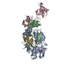
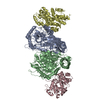
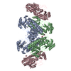
| Title | Crystal Structure of Clostridium perfringens beta-glucuronidase bound with a novel, potent inhibitor 4-(8-(piperazin-1-yl)-1,2,3,4-tetrahydro-[1,2,3]triazino[4',5':4,5]thieno[2,3-c]isoquinolin-5-yl)morpholine | ||||||
 Components Components |
| ||||||
 Keywords Keywords | HYDROLASE/HYDROLASE INHIBITOR / HYDROLASE-HYDROLASE-INHIBITOR complex / HYDROLASE-HYDROLASE INHIBITOR complex | ||||||
| Function / homology |  Function and homology information Function and homology informationbeta-glucuronidase / beta-galactosidase activity / detection of maltose stimulus / maltose transport complex / carbohydrate transport / carbohydrate transmembrane transporter activity / maltose binding / maltose transport / maltodextrin transmembrane transport / ATP-binding cassette (ABC) transporter complex, substrate-binding subunit-containing ...beta-glucuronidase / beta-galactosidase activity / detection of maltose stimulus / maltose transport complex / carbohydrate transport / carbohydrate transmembrane transporter activity / maltose binding / maltose transport / maltodextrin transmembrane transport / ATP-binding cassette (ABC) transporter complex, substrate-binding subunit-containing / ATP-binding cassette (ABC) transporter complex / cell chemotaxis / outer membrane-bounded periplasmic space / carbohydrate metabolic process / periplasmic space / DNA damage response / membrane Similarity search - Function | ||||||
| Biological species |   Clostridium perfringens (bacteria) Clostridium perfringens (bacteria) | ||||||
| Method |  X-RAY DIFFRACTION / SYNCHROTRON / MOLECULAR REPLACEMENT / Resolution: 2.8 Å X-RAY DIFFRACTION / SYNCHROTRON / MOLECULAR REPLACEMENT / Resolution: 2.8 Å | ||||||
 Authors Authors | Wallace, B.D. / Redinbo, M.R. | ||||||
 Citation Citation | Journal: Proc.Natl.Acad.Sci.USA / Year: 2020 Title: Targeted inhibition of gut bacterial beta-glucuronidase activity enhances anticancer drug efficacy. Authors: Bhatt, A.P. / Pellock, S.J. / Biernat, K.A. / Walton, W.G. / Wallace, B.D. / Creekmore, B.C. / Letertre, M.M. / Swann, J.R. / Wilson, I.D. / Roques, J.R. / Darr, D.B. / Bailey, S.T. / ...Authors: Bhatt, A.P. / Pellock, S.J. / Biernat, K.A. / Walton, W.G. / Wallace, B.D. / Creekmore, B.C. / Letertre, M.M. / Swann, J.R. / Wilson, I.D. / Roques, J.R. / Darr, D.B. / Bailey, S.T. / Montgomery, S.A. / Roach, J.M. / Azcarate-Peril, M.A. / Sartor, R.B. / Gharaibeh, R.Z. / Bultman, S.J. / Redinbo, M.R. | ||||||
| History |
|
- Structure visualization
| Structure viewer | Molecule: MolmilJmol/JSmol |
|---|
- Downloads & links
-Download
| PDBx/mmCIF format | 6cxs.cif.gz | 374.2 KB | Display | PDBx/mmCIF format |
|---|---|---|---|---|
| PDB format | pdb6cxs.ent.gz | 298.4 KB | Display | PDB format |
| PDBx/mmJSON format | 6cxs.json.gz | Tree view | PDBx/mmJSON format | |
| Others |  Other downloads Other downloads |
-Validation report
| Summary document | 6cxs_validation.pdf.gz | 1.1 MB | Display | wwPDB validaton report |
|---|---|---|---|---|
| Full document | 6cxs_full_validation.pdf.gz | 1.1 MB | Display | |
| Data in XML | 6cxs_validation.xml.gz | 68 KB | Display | |
| Data in CIF | 6cxs_validation.cif.gz | 92.9 KB | Display | |
| Arichive directory | https://data.pdbj.org/pub/pdb/validation_reports/cx/6cxsftp://data.pdbj.org/pub/pdb/validation_reports/cx/6cxs | HTTPS FTP |
-Related structure data
| Related structure data | 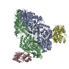 4jkmS S: Starting model for refinement |
|---|---|
| Similar structure data |


-Links
 PDBj
PDBj


- Assembly
| Deposited unit | 
| |||||||||
|---|---|---|---|---|---|---|---|---|---|---|
| 1 |
| |||||||||
| 2 | 
| |||||||||
| Unit cell |
| |||||||||
| Components on special symmetry positions |
|
-Components
| #1: Protein | Mass: 69087.125 Da / Num. of mol.: 2 Source method: isolated from a genetically manipulated source Source: (gene. exp.) Clostridium perfringens (strain 13 / Type A) (bacteria)Strain: 13 / Type A / Gene: bglR / Production host: #2: Protein | Mass: 43993.242 Da / Num. of mol.: 2 / Fragment: residues 27-392 Source method: isolated from a genetically manipulated source Source: (gene. exp.) #3: Chemical |   Mass: 411.524 Da / Num. of mol.: 2 / Source method: obtained synthetically / Formula: C20H25N7OS Mass: 411.524 Da / Num. of mol.: 2 / Source method: obtained synthetically / Formula: C20H25N7OS#4: Water | ChemComp-HOH / |  Mass: 18.015 Da / Num. of mol.: 345 / Source method: isolated from a natural source / Formula: H2O Mass: 18.015 Da / Num. of mol.: 345 / Source method: isolated from a natural source / Formula: H2O |
|---|
-Experimental details
-Experiment
| Experiment | Method: X-RAY DIFFRACTION / Number of used crystals: 1 |
|---|
- Sample preparation
| Crystal | Density Matthews: 3.02 Å3/Da / Density % sol: 59.29 % |
|---|---|
| Crystal grow | Temperature: 289 K / Method: vapor diffusion, hanging drop / Details: 0.1 M MES, 28-36% PEG 400 / PH range: 6.0-6.5 |
-Data collection
| Diffraction | Mean temperature: 100 K | |||||||||||||||||||||||||||||||||||||||||||||||||||||||||||||||||||||||||||||||||||||||||||||||||||||||||||||||||||||||||
|---|---|---|---|---|---|---|---|---|---|---|---|---|---|---|---|---|---|---|---|---|---|---|---|---|---|---|---|---|---|---|---|---|---|---|---|---|---|---|---|---|---|---|---|---|---|---|---|---|---|---|---|---|---|---|---|---|---|---|---|---|---|---|---|---|---|---|---|---|---|---|---|---|---|---|---|---|---|---|---|---|---|---|---|---|---|---|---|---|---|---|---|---|---|---|---|---|---|---|---|---|---|---|---|---|---|---|---|---|---|---|---|---|---|---|---|---|---|---|---|---|---|---|
| Diffraction source | Source: SYNCHROTRON / Site: APS  / Beamline: 23-ID-D / Wavelength: 1 Å / Beamline: 23-ID-D / Wavelength: 1 Å | |||||||||||||||||||||||||||||||||||||||||||||||||||||||||||||||||||||||||||||||||||||||||||||||||||||||||||||||||||||||||
| Detector | Type: MAR scanner 300 mm plate / Detector: IMAGE PLATE / Date: Aug 8, 2012 | |||||||||||||||||||||||||||||||||||||||||||||||||||||||||||||||||||||||||||||||||||||||||||||||||||||||||||||||||||||||||
| Radiation | Protocol: SINGLE WAVELENGTH / Monochromatic (M) / Laue (L): M / Scattering type: x-ray | |||||||||||||||||||||||||||||||||||||||||||||||||||||||||||||||||||||||||||||||||||||||||||||||||||||||||||||||||||||||||
| Radiation wavelength | Wavelength: 1 Å / Relative weight: 1 | |||||||||||||||||||||||||||||||||||||||||||||||||||||||||||||||||||||||||||||||||||||||||||||||||||||||||||||||||||||||||
| Reflection | Resolution: 2.688→48.682 Å / Num. all: 69596 / Num. obs: 69596 / % possible obs: 99.1 % / Redundancy: 4.7 % / Biso Wilson estimate: 60.45 Å2 / Rpim(I) all: 0.05 / Rrim(I) all: 0.114 / Rsym value: 0.101 / Net I/av σ(I): 6.4 / Net I/σ(I): 11.1 / Num. measured all: 327990 | |||||||||||||||||||||||||||||||||||||||||||||||||||||||||||||||||||||||||||||||||||||||||||||||||||||||||||||||||||||||||
| Reflection shell | Diffraction-ID: 1
|
- Processing
| Software |
| |||||||||||||||||||||||||||||||||||||||||||||||||||||||||||||||||||||||||||||||||||||||||||||||||||||||||||||||||||||||||||||||||||||||||||||||||||||||||||||||||
|---|---|---|---|---|---|---|---|---|---|---|---|---|---|---|---|---|---|---|---|---|---|---|---|---|---|---|---|---|---|---|---|---|---|---|---|---|---|---|---|---|---|---|---|---|---|---|---|---|---|---|---|---|---|---|---|---|---|---|---|---|---|---|---|---|---|---|---|---|---|---|---|---|---|---|---|---|---|---|---|---|---|---|---|---|---|---|---|---|---|---|---|---|---|---|---|---|---|---|---|---|---|---|---|---|---|---|---|---|---|---|---|---|---|---|---|---|---|---|---|---|---|---|---|---|---|---|---|---|---|---|---|---|---|---|---|---|---|---|---|---|---|---|---|---|---|---|---|---|---|---|---|---|---|---|---|---|---|---|---|---|---|---|
| Refinement | Method to determine structure: MOLECULAR REPLACEMENT Starting model: 4JKM Resolution: 2.8→48.682 Å / SU ML: 0.34 / Cross valid method: THROUGHOUT / σ(F): 1.33 / Phase error: 23.16 / Stereochemistry target values: ML
| |||||||||||||||||||||||||||||||||||||||||||||||||||||||||||||||||||||||||||||||||||||||||||||||||||||||||||||||||||||||||||||||||||||||||||||||||||||||||||||||||
| Solvent computation | Shrinkage radii: 0.9 Å / VDW probe radii: 1.11 Å / Solvent model: FLAT BULK SOLVENT MODEL | |||||||||||||||||||||||||||||||||||||||||||||||||||||||||||||||||||||||||||||||||||||||||||||||||||||||||||||||||||||||||||||||||||||||||||||||||||||||||||||||||
| Displacement parameters | Biso max: 156.55 Å2 / Biso mean: 61.0749 Å2 / Biso min: 21.24 Å2 | |||||||||||||||||||||||||||||||||||||||||||||||||||||||||||||||||||||||||||||||||||||||||||||||||||||||||||||||||||||||||||||||||||||||||||||||||||||||||||||||||
| Refinement step | Cycle: final / Resolution: 2.8→48.682 Å
| |||||||||||||||||||||||||||||||||||||||||||||||||||||||||||||||||||||||||||||||||||||||||||||||||||||||||||||||||||||||||||||||||||||||||||||||||||||||||||||||||
| LS refinement shell | Refine-ID: X-RAY DIFFRACTION / Rfactor Rfree error: 0 / Total num. of bins used: 22
|
