Movie
Movie Controller


+ Open data

- Basic information
| Entry | 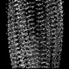 | |||||||||
|---|---|---|---|---|---|---|---|---|---|---|
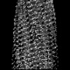
| Title | G175S PMEL CAF amyloid - in vitro polymerized | |||||||||
 Map data Map data | G175S mutant PMEL CAF amyloid, in vitro polymerized | |||||||||
 Sample Sample |
| |||||||||
 Keywords Keywords | melanosome / melanoma / pigment / melanin / amyloid / glaucoma / STRUCTURAL PROTEIN | |||||||||
| Function / homology |  Function and homology information Function and homology informationcis-Golgi network membrane / positive regulation of melanin biosynthetic process / melanin biosynthetic process / melanosome membrane / melanosome organization / multivesicular body membrane / multivesicular body, internal vesicle / Regulation of MITF-M-dependent genes involved in pigmentation / melanosome / endoplasmic reticulum membrane ...cis-Golgi network membrane / positive regulation of melanin biosynthetic process / melanin biosynthetic process / melanosome membrane / melanosome organization / multivesicular body membrane / multivesicular body, internal vesicle / Regulation of MITF-M-dependent genes involved in pigmentation / melanosome / endoplasmic reticulum membrane / Golgi apparatus / endoplasmic reticulum / extracellular exosome / identical protein binding / plasma membrane Similarity search - Function | |||||||||
| Biological species |  Homo sapiens (human) Homo sapiens (human) | |||||||||
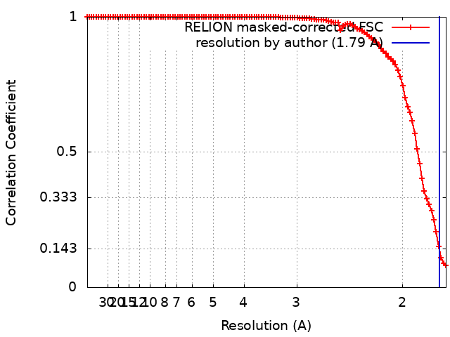
| Method | single particle reconstruction / cryo EM / Resolution: 1.79 Å | |||||||||
 Authors Authors | Oda T / Yanagisawa H | |||||||||
| Funding support |  Japan, Japan,  France, 2 items France, 2 items
| |||||||||
 Citation Citation | Journal: To Be Published Title: Cryo-EM of PMEL Amyloids Reveals Pathogenic Mechanism of G175S in Pigment Dispersion Syndrome. Authors: Yanagisawa H / Oda T | |||||||||
| History |
|
- Structure visualization
| Supplemental images |
|---|
- Downloads & links
-EMDB archive
| Map data | emd_61786.map.gz | 2.6 MB | EMDB map data format | |
|---|---|---|---|---|
| Header (meta data) | emd-61786-v30.xmlemd-61786.xml | 15.4 KB 15.4 KB | Display Display | EMDB header |
| FSC (resolution estimation) | emd_61786_fsc.xml | 10.5 KB | Display | FSC data file |
| Images |  emd_61786.png emd_61786.png | 87.3 KB | ||
| Masks | emd_61786_msk_1.map | 2.8 MB | Mask map | |
| Filedesc metadata | emd-61786.cif.gz | 5.3 KB | ||
| Others | emd_61786_half_map_1.map.gzemd_61786_half_map_2.map.gz | 2.6 MB 2.6 MB | ||
| Archive directory |  http://ftp.pdbj.org/pub/emdb/structures/EMD-61786ftp://ftp.pdbj.org/pub/emdb/structures/EMD-61786 http://ftp.pdbj.org/pub/emdb/structures/EMD-61786ftp://ftp.pdbj.org/pub/emdb/structures/EMD-61786 | HTTPS FTP |
-Validation report
| Summary document | emd_61786_validation.pdf.gz | 1 MB | Display | EMDB validaton report |
|---|---|---|---|---|
| Full document | emd_61786_full_validation.pdf.gz | 1 MB | Display | |
| Data in XML | emd_61786_validation.xml.gz | 12.3 KB | Display | |
| Data in CIF | emd_61786_validation.cif.gz | 16.6 KB | Display | |
| Arichive directory | https://ftp.pdbj.org/pub/emdb/validation_reports/EMD-61786ftp://ftp.pdbj.org/pub/emdb/validation_reports/EMD-61786 | HTTPS FTP |
-Related structure data
| Related structure data | 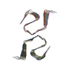 9jsxMC  9jstC  9jsuC  9jsvC  9jswC C: citing same article ( M: atomic model generated by this map |
|---|---|
| Similar structure data |
-Links
| EMDB pages | EMDB (EBI/PDBe) / EMDataResource |
|---|
-Map
| File | Download / File: emd_61786.map.gz / Format: CCP4 / Size: 2.8 MB / Type: IMAGE STORED AS FLOATING POINT NUMBER (4 BYTES) | ||||||||||||||||||||||||||||||||||||
|---|---|---|---|---|---|---|---|---|---|---|---|---|---|---|---|---|---|---|---|---|---|---|---|---|---|---|---|---|---|---|---|---|---|---|---|---|---|
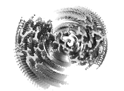
| Annotation | G175S mutant PMEL CAF amyloid, in vitro polymerized | ||||||||||||||||||||||||||||||||||||
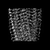
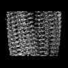
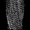
| Projections & slices | Image control
Images are generated by Spider. | ||||||||||||||||||||||||||||||||||||
| Voxel size | X=Y=Z: 0.8784 Å | ||||||||||||||||||||||||||||||||||||
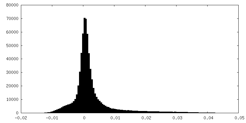
| Density |
| ||||||||||||||||||||||||||||||||||||
| Symmetry | Space group: 1 | ||||||||||||||||||||||||||||||||||||
| Details | EMDB XML:
|
 Z (Sec.)
 Y (Row.)
 X (Col.)


-Supplemental data
-Mask #1
| File | emd_61786_msk_1.map | ||||||||||||
|---|---|---|---|---|---|---|---|---|---|---|---|---|---|
| Projections & Slices |
| ||||||||||||
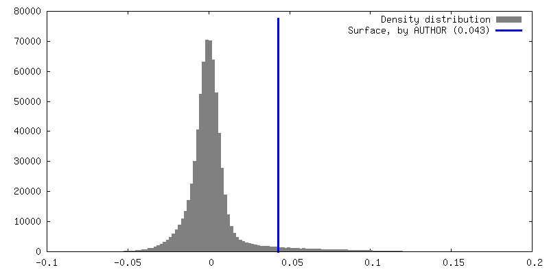
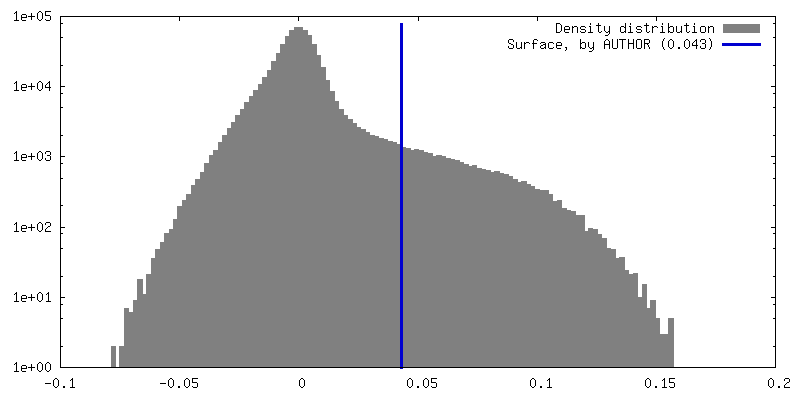
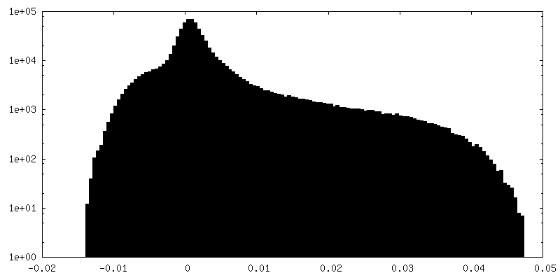
| Density Histograms |


-Half map: Half map B
| File | emd_61786_half_map_1.map | ||||||||||||
|---|---|---|---|---|---|---|---|---|---|---|---|---|---|
| Annotation | Half map B | ||||||||||||
| Projections & Slices |
| ||||||||||||
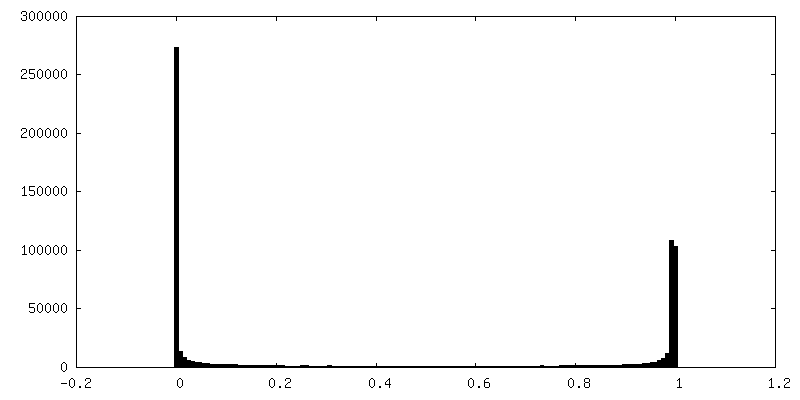
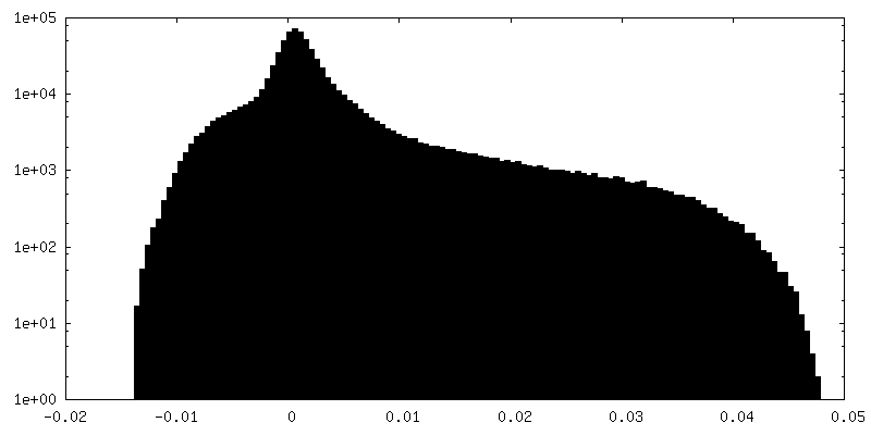
| Density Histograms |


-Half map: Half map A
| File | emd_61786_half_map_2.map | ||||||||||||
|---|---|---|---|---|---|---|---|---|---|---|---|---|---|
| Annotation | Half map A | ||||||||||||
| Projections & Slices |
| ||||||||||||
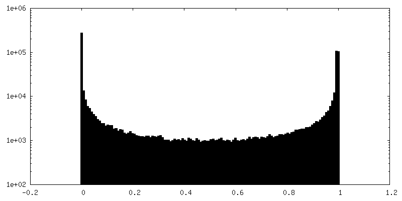
| Density Histograms |


- Sample components
-Entire : G175S mutant PMEL CAF domain, expressed in E. coli
| Entire | Name: G175S mutant PMEL CAF domain, expressed in E. coli |
|---|---|
| Components |
|
-Supramolecule #1: G175S mutant PMEL CAF domain, expressed in E. coli
| Supramolecule | Name: G175S mutant PMEL CAF domain, expressed in E. coli / type: cell / ID: 1 / Parent: 0 / Macromolecule list: all |
|---|---|
| Source (natural) | Organism: Homo sapiens (human) |
-Macromolecule #1: M-alpha
| Macromolecule | Name: M-alpha / type: protein_or_peptide / ID: 1 / Number of copies: 8 / Enantiomer: LEVO |
|---|---|
| Source (natural) | Organism: Homo sapiens (human) |
| Molecular weight | Theoretical: 3.786343 KDa |
| Recombinant expression | Organism:  |
| Sequence | String: FVYVWKTWGQ YWQVLGGPVS GLSIGTSRAM LGTH UniProtKB: Melanocyte protein PMEL |
-Experimental details
-Structure determination
| Method | cryo EM |
|---|---|
 Processing Processing | single particle reconstruction |
| Aggregation state | filament |
-Sample preparation
| Concentration | 0.3 mg/mL |
|---|---|
| Buffer | pH: 4.4 / Component - Concentration: 150.0 mM / Component - Formula: CH3COONa / Component - Name: Sodium acetate |
| Grid | Model: UltrAuFoil R1.2/1.3 / Material: GOLD / Mesh: 300 / Support film - Material: GOLD / Support film - topology: HOLEY |
| Vitrification | Cryogen name: ETHANE / Chamber humidity: 100 % / Chamber temperature: 283 K / Instrument: FEI VITROBOT MARK IV |
| Details | This sample was monodisperse. |
- Electron microscopy
| Microscope | JEOL CRYO ARM 300 |
|---|---|
| Specialist optics | Energy filter - Name: In-column Omega Filter / Energy filter - Slit width: 20 eV |
| Image recording | Film or detector model: GATAN K3 (6k x 4k) / Average exposure time: 5.5 sec. / Average electron dose: 50.0 e/Å2 |
| Electron beam | Acceleration voltage: 300 kV / Electron source:  FIELD EMISSION GUN FIELD EMISSION GUN |
| Electron optics | Illumination mode: FLOOD BEAM / Imaging mode: BRIGHT FIELD / Cs: 2.7 mm / Nominal defocus max: 2.0 µm / Nominal defocus min: 0.5 µm / Nominal magnification: 60000 |
| Sample stage | Cooling holder cryogen: NITROGEN |