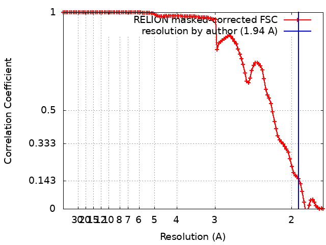
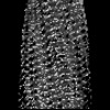
Journal: Nat Commun / Year: 2025 Title: Cryo-EM of wild-type and mutant PMEL amyloid cores reveals structural mechanism of pigment dispersion syndrome. Authors: Haruaki Yanagisawa / Harumi Arai / Tony Wang / Hideyuki Miyazawa / Masahide Kikkawa / Toshiyuki Oda / Abstract: PMEL amyloids serve as essential scaffolds for melanin deposition in melanosomes, playing a crucial role in pigmentation. Despite their importance, the high-resolution structure of PMEL amyloids has ...PMEL amyloids serve as essential scaffolds for melanin deposition in melanosomes, playing a crucial role in pigmentation. Despite their importance, the high-resolution structure of PMEL amyloids has remained unresolved. Using cryo-electron microscopy, we determine near-atomic resolution structures of wild-type PMEL amyloid core, revealing two distinct polymorphic forms with structural features. We further investigate the pathogenic G175S mutation associated with pigment dispersion syndrome (PDS). Structural analysis reveales that G175S introduces an additional hydrogen bond, stabilizing an alternative fibril conformation. In vitro, the G175S mutant exhibits a fourfold increase in polymerization efficiency compared to the wild type. In cells, G175S expression resultes in a twofold increase in intracellular amyloid content and a ~70% increase in extracellular amyloids, without altering melanosome morphology or number. These results indicate that the G175S mutation enhances amyloidogenesis within melanosomes, elevating amyloid load and potentially contributing to PDS pathophysiology. This study provides molecular insights into PMEL amyloid formation, highlighting its structural diversity and dysregulation in pigmentation disorders.
In the structure databanks used in Yorodumi, some data are registered as the other names, "COVID-19 virus" and "2019-nCoV". Here are the details of the virus and the list of structure data.
Jan 31, 2019. EMDB accession codes are about to change! (news from PDBe EMDB page)
EMDB accession codes are about to change! (news from PDBe EMDB page)
The allocation of 4 digits for EMDB accession codes will soon come to an end. Whilst these codes will remain in use, new EMDB accession codes will include an additional digit and will expand incrementally as the available range of codes is exhausted. The current 4-digit format prefixed with “EMD-” (i.e. EMD-XXXX) will advance to a 5-digit format (i.e. EMD-XXXXX), and so on. It is currently estimated that the 4-digit codes will be depleted around Spring 2019, at which point the 5-digit format will come into force.
The EM Navigator/Yorodumi systems omit the EMD- prefix.
Related info.:Q: What is EMD? / ID/Accession-code notation in Yorodumi/EM Navigator
Yorodumi is a browser for structure data from EMDB, PDB, SASBDB, etc.
This page is also the successor to EM Navigator detail page, and also detail information page/front-end page for Omokage search.
The word "yorodu" (or yorozu) is an old Japanese word meaning "ten thousand". "mi" (miru) is to see.
Related info.:EMDB / PDB / SASBDB / Comparison of 3 databanks / Yorodumi Search / Aug 31, 2016. New EM Navigator & Yorodumi / Yorodumi Papers / Jmol/JSmol / Function and homology information / Changes in new EM Navigator and Yorodumi
 Movie
Movie Controller


 Open data
 Basic information
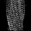
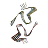
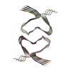
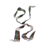
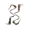
 Map data
 Sample
 Keywords
 Function and homology information
 Homo sapiens (human)
 Authors
 Japan,
  France, 2 items
  Citation
 Structure visualization
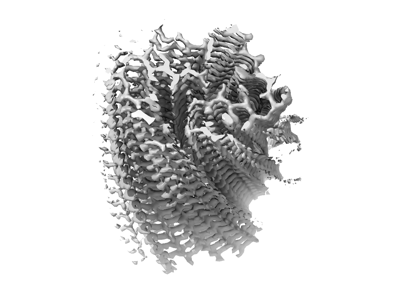
 Downloads & links
 emd_61785.png
 http://ftp.pdbj.org/pub/emdb/structures/EMD-61785
 Z (Sec.)
 Y (Row.)
 X (Col.)


 Sample components
 Processing
 Electron microscopy
 FIELD EMISSION GUN