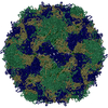
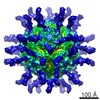
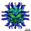
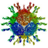
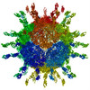
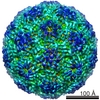
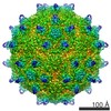
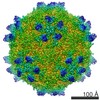
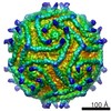
ジャーナル: J Virol / 年: 2015 タイトル: Nectin-like interactions between poliovirus and its receptor trigger conformational changes associated with cell entry. 著者: Mike Strauss / David J Filman / David M Belnap / Naiqian Cheng / Roane T Noel / James M Hogle / 要旨: Poliovirus infection is initiated by attachment to a receptor on the cell surface called Pvr or CD155. At physiological temperatures, the receptor catalyzes an irreversible expansion of the virus to ...Poliovirus infection is initiated by attachment to a receptor on the cell surface called Pvr or CD155. At physiological temperatures, the receptor catalyzes an irreversible expansion of the virus to form an expanded form of the capsid called the 135S particle. This expansion results in the externalization of the myristoylated capsid protein VP4 and the N-terminal extension of the capsid protein VP1, both of which become inserted into the cell membrane. Structures of the expanded forms of poliovirus and of several related viruses have recently been reported. However, until now, it has been unclear how receptor binding triggers viral expansion at physiological temperature. Here, we report poliovirus in complex with an enzymatically partially deglycosylated form of the 3-domain ectodomain of Pvr at a 4-Å resolution, as determined by cryo-electron microscopy. The interaction of the receptor with the virus in this structure is reminiscent of the interactions of Pvr with its natural ligands. At a low temperature, the receptor induces very few changes in the structure of the virus, with the largest changes occurring within the footprint of the receptor, and in a loop of the internal protein VP4. Changes in the vicinity of the receptor include the displacement of a natural lipid ligand (called "pocket factor"), demonstrating that the loss of this ligand, alone, is not sufficient to induce particle expansion. Finally, analogies with naturally occurring ligand binding in the nectin family suggest which specific structural rearrangements in the virus-receptor complex could help to trigger the irreversible expansion of the capsid. IMPORTANCE: The cell-surface receptor (Pvr) catalyzes a large structural change in the virus that exposes membrane-binding protein chains. We fitted known atomic models of the virus and Pvr into ...IMPORTANCE: The cell-surface receptor (Pvr) catalyzes a large structural change in the virus that exposes membrane-binding protein chains. We fitted known atomic models of the virus and Pvr into three-dimensional experimental maps of the receptor-virus complex. The molecular interactions we see between poliovirus and its receptor are reminiscent of the nectin family, by involving the burying of otherwise-exposed hydrophobic groups. Importantly, poliovirus expansion is regulated by the binding of a lipid molecule within the viral capsid. We show that receptor binding either causes this molecule to be expelled or requires it, but that its loss is not sufficient to trigger irreversible expansion. Based on our model, we propose testable hypotheses to explain how the viral shell becomes destabilized, leading to RNA uncoating. These findings give us a better understanding of how poliovirus has evolved to exploit a natural process of its host to penetrate the membrane barrier.
解像度のタイプ: BY AUTHOR / 解像度: 4.0 Å / 解像度の算出法: OTHER / ソフトウェア - 名称: Relion1.2, GeFrealign / 詳細: This map has a B-factor of -60 applied to it. / 使用した粒子像数: 9248
After rigid-body fitting, manual model building was alternated with automated stereochemically restrained refinement, minimizing the discrepancy between experimental and model-based Fourier amplitudes and phases.
精密化
空間: RECIPROCAL / プロトコル: RIGID BODY FIT / 当てはまり具合の基準: pseudo-real space
After rigid-body fitting, manual model building was alternated with automated stereochemically restrained refinement, minimizing the discrepancy between experimental and model-based Fourier amplitudes and phases.
精密化
空間: RECIPROCAL / プロトコル: FLEXIBLE FIT / 当てはまり具合の基準: pseudo-real space
 ムービー
ムービー コントローラー


 データを開く
 基本情報
基本情報 マップデータ
 試料
試料 キーワード
キーワード 機能・相同性情報
 Homo sapiens (ヒト) /
 Human poliovirus 1 Mahoney (ポリオウイルス)
 データ登録者
 引用
引用
 構造の表示
構造の表示
 ダウンロードとリンク
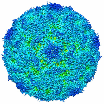 400_6148.gif
 80_6148.gif
 http://ftp.pdbj.org/pub/emdb/structures/EMD-6148
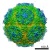


 試料の構成要素
 解析
解析 電子顕微鏡法
 FIELD EMISSION GUN