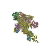
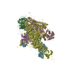
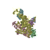
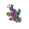
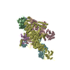
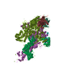
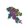
Journal: EMBO Rep / Year: 2019 Title: High-resolution archaellum structure reveals a conserved metal-binding site. Authors: Vladimir A Meshcheryakov / Satoshi Shibata / Makoto Tokoro Schreiber / Alejandro Villar-Briones / Kenneth F Jarrell / Shin-Ichi Aizawa / Matthias Wolf / Abstract: Many archaea swim by means of archaella. While the archaellum is similar in function to its bacterial counterpart, its structure, composition, and evolution are fundamentally different. Archaella are ...Many archaea swim by means of archaella. While the archaellum is similar in function to its bacterial counterpart, its structure, composition, and evolution are fundamentally different. Archaella are related to archaeal and bacterial type IV pili. Despite recent advances, our understanding of molecular processes governing archaellum assembly and stability is still incomplete. Here, we determine the structures of archaella by X-ray crystallography and cryo-EM The crystal structure of FlaB1 is the first and only crystal structure of any archaellin to date at a resolution of 1.5 Å, which is put into biological context by a cryo-EM reconstruction from archaella at 4 Å resolution created with helical single-particle analysis. Our results indicate that the archaellum is predominantly composed of FlaB1. We identify N-linked glycosylation by cryo-EM and mass spectrometry. The crystal structure reveals a highly conserved metal-binding site, which is validated by mass spectrometry and electron energy-loss spectroscopy. We show that the metal-binding site, which appears to be a widespread property of archaellin, is required for filament integrity.
Cryogen: NITROGEN / Specimen holder model: FEI TITAN KRIOS AUTOGRID HOLDER / Temperature (max): 100 K / Temperature (min): 77 K / Residual tilt: 0.1 mradians
Image recording
Average exposure time: 12 sec. / Electron dose: 96 e/Å2 / Detector mode: SUPER-RESOLUTION / Film or detector model: GATAN K2 SUMMIT (4k x 4k) / Num. of grids imaged: 1 / Num. of real images: 2000
EM imaging optics
Energyfilter name: GIF Quantum LS / Energyfilter upper: 20 eV / Energyfilter lower: 0 eV
Image scans
Sampling size: 5 µm / Width: 7676 / Height: 7420 / Movie frames/image: 4 / Used frames/image: 1-48
Resolution: 4 Å / Resolution method: FSC 0.143 CUT-OFF / Num. of particles: 110747 / Algorithm: FOURIER SPACE / Num. of class averages: 100 / Symmetry type: POINT
Atomic model building
Protocol: FLEXIBLE FIT / Space: REAL
Refine LS restraints
Refine-ID
Type
Dev ideal
Number
ELECTRONMICROSCOPY
f_bond_d
0.004
25848
ELECTRONMICROSCOPY
f_angle_d
0.969
35244
ELECTRONMICROSCOPY
f_dihedral_angle_d
4.358
15318
ELECTRONMICROSCOPY
f_chiral_restr
0.069
4464
ELECTRONMICROSCOPY
f_plane_restr
0.006
4554
+
About Yorodumi
-
News
-
Feb 9, 2022. New format data for meta-information of EMDB entries
New format data for meta-information of EMDB entries
Version 3 of the EMDB header file is now the official format.
The previous official version 1.9 will be removed from the archive.
In the structure databanks used in Yorodumi, some data are registered as the other names, "COVID-19 virus" and "2019-nCoV". Here are the details of the virus and the list of structure data.
Jan 31, 2019. EMDB accession codes are about to change! (news from PDBe EMDB page)
EMDB accession codes are about to change! (news from PDBe EMDB page)
The allocation of 4 digits for EMDB accession codes will soon come to an end. Whilst these codes will remain in use, new EMDB accession codes will include an additional digit and will expand incrementally as the available range of codes is exhausted. The current 4-digit format prefixed with “EMD-” (i.e. EMD-XXXX) will advance to a 5-digit format (i.e. EMD-XXXXX), and so on. It is currently estimated that the 4-digit codes will be depleted around Spring 2019, at which point the 5-digit format will come into force.
The EM Navigator/Yorodumi systems omit the EMD- prefix.
Related info.:Q: What is EMD? / ID/Accession-code notation in Yorodumi/EM Navigator
Yorodumi is a browser for structure data from EMDB, PDB, SASBDB, etc.
This page is also the successor to EM Navigator detail page, and also detail information page/front-end page for Omokage search.
The word "yorodu" (or yorozu) is an old Japanese word meaning "ten thousand". "mi" (miru) is to see.
Related info.:EMDB / PDB / SASBDB / Comparison of 3 databanks / Yorodumi Search / Aug 31, 2016. New EM Navigator & Yorodumi / Yorodumi Papers / Jmol/JSmol / Function and homology information / Changes in new EM Navigator and Yorodumi
 Movie
Movie Controller


 Open data
 Basic information
 Components
 Keywords
 Function and homology information
 Methanococcus maripaludis (archaea)
 Authors
 Japan, 2items
  Citation
 Structure visualization
 Downloads & links
 Other downloads
 PDBj
PDBj Assembly
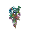
 Sample preparation
 Electron microscopy imaging
 FIELD EMISSION GUN / Accelerating voltage: 300 kV / Illumination mode: FLOOD BEAM
 Processing