+ Open data
- Basic information
| Entry | Database: PDB / ID: 5yud | ||||||||||||||||||||||||||||||||||||
|---|---|---|---|---|---|---|---|---|---|---|---|---|---|---|---|---|---|---|---|---|---|---|---|---|---|---|---|---|---|---|---|---|---|---|---|---|---|
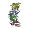
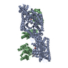
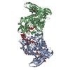
| Title | Flagellin derivative in complex with the NLR protein NAIP5 | ||||||||||||||||||||||||||||||||||||
 Components Components |
| ||||||||||||||||||||||||||||||||||||
 Keywords Keywords | IMMUNE SYSTEM / Flagellin / NAIP5 / NLRC4 / Cryo-EM | ||||||||||||||||||||||||||||||||||||
| Function / homology |  Function and homology information Function and homology informationTLR5 cascade / MyD88 cascade initiated on plasma membrane / NFkB and MAPK activation mediated by TRAF6 / IPAF inflammasome complex / The IPAF inflammasome / bacterial-type flagellum / cysteine-type endopeptidase inhibitor activity involved in apoptotic process / pyroptotic inflammatory response / detection of bacterium / positive regulation of interleukin-1 beta production ...TLR5 cascade / MyD88 cascade initiated on plasma membrane / NFkB and MAPK activation mediated by TRAF6 / IPAF inflammasome complex / The IPAF inflammasome / bacterial-type flagellum / cysteine-type endopeptidase inhibitor activity involved in apoptotic process / pyroptotic inflammatory response / detection of bacterium / positive regulation of interleukin-1 beta production / defense response to Gram-negative bacterium / defense response to bacterium / inflammatory response / receptor ligand activity / symbiont entry into host cell / innate immune response / apoptotic process / negative regulation of apoptotic process / structural molecule activity / extracellular space / extracellular region / ATP binding / metal ion binding Similarity search - Function | ||||||||||||||||||||||||||||||||||||
| Biological species |   Salmonella typhimurium (bacteria) Salmonella typhimurium (bacteria) | ||||||||||||||||||||||||||||||||||||
| Method | ELECTRON MICROSCOPY / single particle reconstruction / cryo EM / Resolution: 4.28 Å | ||||||||||||||||||||||||||||||||||||
 Authors Authors | Yang, X.R. / Yang, F. / Wang, W.G. / Lin, G.Z. | ||||||||||||||||||||||||||||||||||||
| Funding support |  China, 6items China, 6items
| ||||||||||||||||||||||||||||||||||||
 Citation Citation |  Journal: Cell Res / Year: 2018 Journal: Cell Res / Year: 2018Title: Structural basis for specific flagellin recognition by the NLR protein NAIP5. Authors: Xinru Yang / Fan Yang / Weiguang Wang / Guangzhong Lin / Zehan Hu / Zhifu Han / Yijun Qi / Liman Zhang / Jiawei Wang / Sen-Fang Sui / Jijie Chai /    Abstract: The nucleotide-binding domain- and leucine-rich repeat (LRR)-containing proteins (NLRs) function as intracellular immune receptors to detect the presence of pathogen- or host-derived signals. The ...The nucleotide-binding domain- and leucine-rich repeat (LRR)-containing proteins (NLRs) function as intracellular immune receptors to detect the presence of pathogen- or host-derived signals. The mechanisms of how NLRs sense their ligands remain elusive. Here we report the structure of a bacterial flagellin derivative in complex with the NLR proteins NAIP5 and NLRC4 determined by cryo-electron microscopy at 4.28 Å resolution. The structure revealed that the flagellin derivative forms two parallel helices interacting with multiple domains including BIR1 and LRR of NAIP5. Binding to NAIP5 results in a nearly complete burial of the flagellin derivative, thus stabilizing the active conformation of NAIP5. The extreme C-terminal side of the flagellin is anchored to a sterically constrained binding pocket of NAIP5, which likely acts as a structural determinant for discrimination of different bacterial flagellins by NAIP5, a notion further supported by biochemical data. Taken together, our results shed light on the molecular mechanisms underlying NLR ligand perception. | ||||||||||||||||||||||||||||||||||||
| History |
|
- Structure visualization
| Movie |
 Movie viewer Movie viewer |
|---|---|
| Structure viewer | Molecule:  Molmil Molmil Jmol/JSmol Jmol/JSmol |
- Downloads & links
- Download
| PDBx/mmCIF format |  5yud.cif.gz 5yud.cif.gz | 255.3 KB | Display |  PDBx/mmCIF format PDBx/mmCIF format |
|---|---|---|---|---|
| PDB format |  pdb5yud.ent.gz pdb5yud.ent.gz | 198.9 KB | Display |  PDB format PDB format |
| PDBx/mmJSON format |  5yud.json.gz 5yud.json.gz | Tree view |  PDBx/mmJSON format PDBx/mmJSON format | |
| Others |  Other downloads Other downloads |
-Validation report
| Summary document |  5yud_validation.pdf.gz 5yud_validation.pdf.gz | 995.5 KB | Display |  wwPDB validaton report wwPDB validaton report |
|---|---|---|---|---|
| Full document |  5yud_full_validation.pdf.gz 5yud_full_validation.pdf.gz | 1.1 MB | Display | |
| Data in XML |  5yud_validation.xml.gz 5yud_validation.xml.gz | 57.6 KB | Display | |
| Data in CIF |  5yud_validation.cif.gz 5yud_validation.cif.gz | 85.1 KB | Display | |
| Arichive directory |  https://data.pdbj.org/pub/pdb/validation_reports/yu/5yud https://data.pdbj.org/pub/pdb/validation_reports/yu/5yud ftp://data.pdbj.org/pub/pdb/validation_reports/yu/5yud ftp://data.pdbj.org/pub/pdb/validation_reports/yu/5yud | HTTPS FTP |
-Related structure data
| Related structure data |  6845MC M: map data used to model this data C: citing same article ( |
|---|---|
| Similar structure data |
- Links
Links
- Assembly
| Deposited unit | 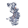
|
|---|---|
| 1 |
|
- Components
| #1: Protein | Mass: 160018.047 Da / Num. of mol.: 1 Source method: isolated from a genetically manipulated source Source: (gene. exp.)   |
|---|---|
| #2: Protein | Mass: 7665.362 Da / Num. of mol.: 1 Source method: isolated from a genetically manipulated source Source: (gene. exp.)  Salmonella typhimurium (strain LT2 / SGSC1412 / ATCC 700720) (bacteria) Salmonella typhimurium (strain LT2 / SGSC1412 / ATCC 700720) (bacteria)Strain: LT2 / SGSC1412 / ATCC 700720 / Gene: fljB, H2, STM2771, fliC, flaF, hag, STM1959 / Production host:  |
| #3: Chemical | ChemComp-ATP / |
| Has protein modification | N |
-Experimental details
-Experiment
| Experiment | Method: ELECTRON MICROSCOPY |
|---|---|
| EM experiment | Aggregation state: PARTICLE / 3D reconstruction method: single particle reconstruction |
- Sample preparation
| Component |
| ||||||||||||||||||||||||
|---|---|---|---|---|---|---|---|---|---|---|---|---|---|---|---|---|---|---|---|---|---|---|---|---|---|
| Source (natural) |
| ||||||||||||||||||||||||
| Source (recombinant) |
| ||||||||||||||||||||||||
| Buffer solution | pH: 8 | ||||||||||||||||||||||||
| Specimen | Embedding applied: NO / Shadowing applied: NO / Staining applied: NO / Vitrification applied: YES | ||||||||||||||||||||||||
| Vitrification | Cryogen name: ETHANE |
- Electron microscopy imaging
| Experimental equipment |  Model: Titan Krios / Image courtesy: FEI Company |
|---|---|
| Microscopy | Model: FEI TITAN KRIOS |
| Electron gun | Electron source:  FIELD EMISSION GUN / Accelerating voltage: 300 kV / Illumination mode: FLOOD BEAM FIELD EMISSION GUN / Accelerating voltage: 300 kV / Illumination mode: FLOOD BEAM |
| Electron lens | Mode: BRIGHT FIELD |
| Image recording | Electron dose: 50 e/Å2 / Film or detector model: GATAN K2 SUMMIT (4k x 4k) |
- Processing
| Software | Name: PHENIX / Version: 1.10.1_2155: / Classification: refinement | ||||||||||||||||||||||||
|---|---|---|---|---|---|---|---|---|---|---|---|---|---|---|---|---|---|---|---|---|---|---|---|---|---|
| EM software | Name: PHENIX / Category: model refinement | ||||||||||||||||||||||||
| CTF correction | Type: PHASE FLIPPING AND AMPLITUDE CORRECTION | ||||||||||||||||||||||||
| 3D reconstruction | Resolution: 4.28 Å / Resolution method: FSC 0.143 CUT-OFF / Num. of particles: 626608 / Symmetry type: POINT | ||||||||||||||||||||||||
| Refine LS restraints |
|
 Movie
Movie Controller




 PDBj
PDBj


