 Movie
Movie Controller


+ Open data

- Basic information
| Entry | Database: PDB / ID: 5yrn | ||||||
|---|---|---|---|---|---|---|---|
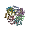
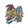
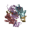
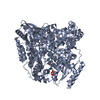
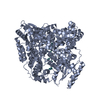
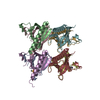
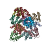
| Title | Structure of RIP2 CARD domain | ||||||
 Components Components | Receptor-interacting serine/threonine-protein kinase 2 | ||||||
 Keywords Keywords | TRANSFERASE / Innate Immune signaling complex / IMMUNE SYSTEM | ||||||
| Function / homology |  Function and homology information Function and homology informationresponse to interleukin-18 / toll-like receptor 2 signaling pathway / positive regulation of T-helper 1 cell differentiation / immature T cell proliferation in thymus / positive regulation of cytokine-mediated signaling pathway / positive regulation of T-helper 1 type immune response / positive regulation of xenophagy / nucleotide-binding oligomerization domain containing 1 signaling pathway / caspase binding / xenophagy ...response to interleukin-18 / toll-like receptor 2 signaling pathway / positive regulation of T-helper 1 cell differentiation / immature T cell proliferation in thymus / positive regulation of cytokine-mediated signaling pathway / positive regulation of T-helper 1 type immune response / positive regulation of xenophagy / nucleotide-binding oligomerization domain containing 1 signaling pathway / caspase binding / xenophagy / LIM domain binding / positive regulation of protein K63-linked ubiquitination / positive regulation of stress-activated MAPK cascade / cellular response to muramyl dipeptide / CD4-positive, alpha-beta T cell proliferation / CARD domain binding / positive regulation of immature T cell proliferation in thymus / cellular response to peptidoglycan / response to interleukin-12 / JUN kinase kinase kinase activity / positive regulation of CD4-positive, alpha-beta T cell proliferation / nucleotide-binding oligomerization domain containing 2 signaling pathway / positive regulation of peptidyl-tyrosine phosphorylation / positive regulation of macrophage cytokine production / toll-like receptor 4 signaling pathway / response to exogenous dsRNA / cellular response to lipoteichoic acid / positive regulation of interferon-alpha production / stress-activated MAPK cascade / positive regulation of interleukin-2 production / ERK1 and ERK2 cascade / JNK cascade / positive regulation of chemokine production / canonical NF-kappaB signal transduction / positive regulation of interleukin-12 production / signaling adaptor activity / positive regulation of interferon-beta production / lipopolysaccharide-mediated signaling pathway / response to interleukin-1 / p75NTR recruits signalling complexes / positive regulation of protein ubiquitination / positive regulation of interleukin-1 beta production / JNK (c-Jun kinases) phosphorylation and activation mediated by activated human TAK1 / non-specific protein-tyrosine kinase / activated TAK1 mediates p38 MAPK activation / non-membrane spanning protein tyrosine kinase activity / NOD1/2 Signaling Pathway / protein homooligomerization / TAK1-dependent IKK and NF-kappa-B activation / positive regulation of interleukin-6 production / positive regulation of JNK cascade / positive regulation of type II interferon production / cytokine-mediated signaling pathway / Interleukin-1 signaling / positive regulation of tumor necrosis factor production / Ovarian tumor domain proteases / Downstream TCR signaling / T cell receptor signaling pathway / vesicle / cytoskeleton / adaptive immune response / positive regulation of canonical NF-kappaB signal transduction / positive regulation of ERK1 and ERK2 cascade / non-specific serine/threonine protein kinase / defense response to bacterium / defense response to Gram-positive bacterium / positive regulation of apoptotic process / inflammatory response / signaling receptor binding / innate immune response / protein serine kinase activity / protein serine/threonine kinase activity / apoptotic process / SARS-CoV-2 activates/modulates innate and adaptive immune responses / endoplasmic reticulum / signal transduction / protein homodimerization activity / positive regulation of transcription by RNA polymerase II / protein-containing complex / ATP binding / identical protein binding / plasma membrane / cytoplasm / cytosol Similarity search - Function | ||||||
| Biological species |  Homo sapiens (human) Homo sapiens (human) | ||||||
| Method | ELECTRON MICROSCOPY / helical reconstruction / cryo EM / Resolution: 4.1 Å | ||||||
 Authors Authors | Wu, B. / Gong, Q. | ||||||
| Funding support |  Singapore, 1items Singapore, 1items
| ||||||
 Citation Citation | Journal: Nat Commun / Year: 2018 Title: Structural basis of RIP2 activation and signaling. Authors: Qin Gong / Ziqi Long / Franklin L Zhong / Daniel Eng Thiam Teo / Yibo Jin / Zhan Yin / Zhao Zhi Boo / Yaming Zhang / Jiawen Zhang / Renliang Yang / Shashi Bhushan / Bruno Reversade / Zongli Li / Bin Wu /     Abstract: Signals arising from bacterial infections are detected by pathogen recognition receptors (PRRs) and are transduced by specialized adapter proteins in mammalian cells. The Receptor-interacting- ...Signals arising from bacterial infections are detected by pathogen recognition receptors (PRRs) and are transduced by specialized adapter proteins in mammalian cells. The Receptor-interacting-serine/threonine-protein kinase 2 (RIPK2 or RIP2) is such an adapter protein that is critical for signal propagation of the Nucleotide-binding-oligomerization-domain-containing proteins 1/2 (NOD1 and NOD2). Dysregulation of this signaling pathway leads to defects in bacterial detection and in some cases autoimmune diseases. Here, we show that the Caspase-activation-and-recruitment-domain (CARD) of RIP2 (RIP2-CARD) forms oligomeric structures upon stimulation by either NOD1-CARD or NOD2-2CARD. We reconstitute this complex, termed the RIPosome in vitro and solve the cryo-EM filament structure of the active RIP2-CARD complex at 4.1 Å resolution. The structure suggests potential mechanisms by which CARD domains from NOD1 and NOD2 initiate the oligomerization process of RIP2-CARD. Together with structure guided mutagenesis experiments at the CARD-CARD interfaces, we demonstrate molecular mechanisms how RIP2 is activated and self-propagating such signal. | ||||||
| History |
|
- Structure visualization
| Movie |
Movie viewer |
|---|---|
| Structure viewer | Molecule: MolmilJmol/JSmol |
- Downloads & links
-Download
| PDBx/mmCIF format | 5yrn.cif.gz | 181.5 KB | Display | PDBx/mmCIF format |
|---|---|---|---|---|
| PDB format | pdb5yrn.ent.gz | 143.1 KB | Display | PDB format |
| PDBx/mmJSON format | 5yrn.json.gz | Tree view | PDBx/mmJSON format | |
| Others |  Other downloads Other downloads |
-Validation report
| Arichive directory | https://data.pdbj.org/pub/pdb/validation_reports/yr/5yrnftp://data.pdbj.org/pub/pdb/validation_reports/yr/5yrn | HTTPS FTP |
|---|
-Related structure data
| Related structure data | 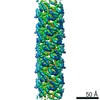 6842MC  6663C C: citing same article ( M: map data used to model this data |
|---|---|
| Similar structure data |

-Links
 PDBj
PDBj


- Assembly
| Deposited unit | 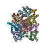
|
|---|---|
| 1 |
|
-Components
| #1: Protein | Mass: 12621.438 Da / Num. of mol.: 12 / Fragment: CARD domain Source method: isolated from a genetically manipulated source Source: (gene. exp.) Homo sapiens (human) / Gene: RIPK2, CARDIAK, RICK, RIP2, UNQ277/PRO314/PRO34092 / Production host:  References: UniProt: O43353, non-specific serine/threonine protein kinase, non-specific protein-tyrosine kinase |
|---|
-Experimental details
-Experiment
| Experiment | Method: ELECTRON MICROSCOPY |
|---|---|
| EM experiment | Aggregation state: FILAMENT / 3D reconstruction method: helical reconstruction |
- Sample preparation
| Component | Name: Active RIP2 signaling complex / Type: ORGANELLE OR CELLULAR COMPONENT / Entity ID: all / Source: RECOMBINANT |
|---|---|
| Source (natural) | Organism: Homo sapiens (human) |
| Source (recombinant) | Organism: |
| Buffer solution | pH: 7.4 |
| Specimen | Conc.: 0.2 mg/ml / Embedding applied: NO / Shadowing applied: NO / Staining applied: NO / Vitrification applied: YES |
| Specimen support | Grid material: COPPER |
| Vitrification | Cryogen name: METHANE |
- Electron microscopy imaging
| Experimental equipment |  Model: Tecnai Polara / Image courtesy: FEI Company |
|---|---|
| Microscopy | Model: FEI POLARA 300 |
| Electron gun | Electron source:  FIELD EMISSION GUN / Accelerating voltage: 300 kV / Illumination mode: FLOOD BEAM FIELD EMISSION GUN / Accelerating voltage: 300 kV / Illumination mode: FLOOD BEAM |
| Electron lens | Mode: BRIGHT FIELD |
| Specimen holder | Specimen holder model: FEI TITAN KRIOS AUTOGRID HOLDER |
| Image recording | Electron dose: 8 e/Å2 / Detector mode: COUNTING / Film or detector model: GATAN K2 SUMMIT (4k x 4k) / Num. of grids imaged: 3 / Num. of real images: 3100 |
- Processing
| Software | Name: PHENIX / Version: dev_2405: / Classification: refinement | ||||||||||||||||||||||||||||||||||||
|---|---|---|---|---|---|---|---|---|---|---|---|---|---|---|---|---|---|---|---|---|---|---|---|---|---|---|---|---|---|---|---|---|---|---|---|---|---|
| EM software |
| ||||||||||||||||||||||||||||||||||||
| CTF correction | Type: PHASE FLIPPING AND AMPLITUDE CORRECTION | ||||||||||||||||||||||||||||||||||||
| Helical symmerty | Angular rotation/subunit: -101.373 ° / Axial rise/subunit: 4.936 Å / Axial symmetry: C1 | ||||||||||||||||||||||||||||||||||||
| Particle selection | Num. of particles selected: 658160 | ||||||||||||||||||||||||||||||||||||
| 3D reconstruction | Resolution: 4.1 Å / Resolution method: FSC 0.143 CUT-OFF / Num. of particles: 142330 Details: Model building was based on a cropped-out density segment. Pseudo-crystallographic refinement showed that the central segment of the map is good until 3.5-3.7 A. Symmetry type: HELICAL | ||||||||||||||||||||||||||||||||||||
| Atomic model building | B value: 202.7 / Protocol: RIGID BODY FIT / Space: RECIPROCAL / Target criteria: Rfree value | ||||||||||||||||||||||||||||||||||||
| Refinement step | Cycle: 1 / Total: 9000 | ||||||||||||||||||||||||||||||||||||
| Refine LS restraints |
|
