 Movie
Movie Controller


+ Open data

- Basic information
| Entry | Database: EMDB / ID: EMD-6663 | |||||||||
|---|---|---|---|---|---|---|---|---|---|---|
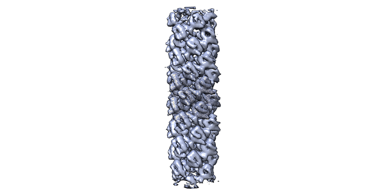
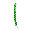
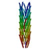
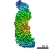
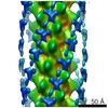
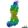
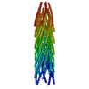
| Title | Structure of RIP2 CARD domain | |||||||||
 Map data Map data | ||||||||||
 Sample Sample |
| |||||||||
| Biological species |  Homo sapiens (human) Homo sapiens (human) | |||||||||
| Method | helical reconstruction / cryo EM / Resolution: 4.85 Å | |||||||||
 Authors Authors | Wu B / Gong Q | |||||||||
 Citation Citation | Journal: Nat Commun / Year: 2018 Title: Structural basis of RIP2 activation and signaling. Authors: Qin Gong / Ziqi Long / Franklin L Zhong / Daniel Eng Thiam Teo / Yibo Jin / Zhan Yin / Zhao Zhi Boo / Yaming Zhang / Jiawen Zhang / Renliang Yang / Shashi Bhushan / Bruno Reversade / Zongli Li / Bin Wu /      Abstract: Signals arising from bacterial infections are detected by pathogen recognition receptors (PRRs) and are transduced by specialized adapter proteins in mammalian cells. The Receptor-interacting- ...Signals arising from bacterial infections are detected by pathogen recognition receptors (PRRs) and are transduced by specialized adapter proteins in mammalian cells. The Receptor-interacting-serine/threonine-protein kinase 2 (RIPK2 or RIP2) is such an adapter protein that is critical for signal propagation of the Nucleotide-binding-oligomerization-domain-containing proteins 1/2 (NOD1 and NOD2). Dysregulation of this signaling pathway leads to defects in bacterial detection and in some cases autoimmune diseases. Here, we show that the Caspase-activation-and-recruitment-domain (CARD) of RIP2 (RIP2-CARD) forms oligomeric structures upon stimulation by either NOD1-CARD or NOD2-2CARD. We reconstitute this complex, termed the RIPosome in vitro and solve the cryo-EM filament structure of the active RIP2-CARD complex at 4.1 Å resolution. The structure suggests potential mechanisms by which CARD domains from NOD1 and NOD2 initiate the oligomerization process of RIP2-CARD. Together with structure guided mutagenesis experiments at the CARD-CARD interfaces, we demonstrate molecular mechanisms how RIP2 is activated and self-propagating such signal. | |||||||||
| History |
|
- Structure visualization
| Movie |
 Movie viewer Movie viewer |
|---|---|
| Structure viewer | EM map: SurfViewMolmilJmol/JSmol |
| Supplemental images |
- Downloads & links
-EMDB archive
| Map data | emd_6663.map.gz | 28 MB | EMDB map data format | |
|---|---|---|---|---|
| Header (meta data) | emd-6663-v30.xmlemd-6663.xml | 13.8 KB 13.8 KB | Display Display | EMDB header |
| Images |  emd_6663.png emd_6663.png | 98 KB | ||
| Others | emd_6663_half_map_1.map.gzemd_6663_half_map_2.map.gz | 23.3 MB 23.3 MB | ||
| Archive directory |  http://ftp.pdbj.org/pub/emdb/structures/EMD-6663ftp://ftp.pdbj.org/pub/emdb/structures/EMD-6663 http://ftp.pdbj.org/pub/emdb/structures/EMD-6663ftp://ftp.pdbj.org/pub/emdb/structures/EMD-6663 | HTTPS FTP |
-Related structure data
| Related structure data |  5h4a 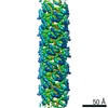 6842C 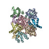 5yrnC M: atomic model generated by this map C: citing same article ( |
|---|---|
| Similar structure data |

-Links
| EMDB pages | EMDB (EBI/PDBe) / EMDataResource |
|---|
-Map
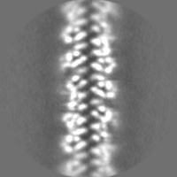
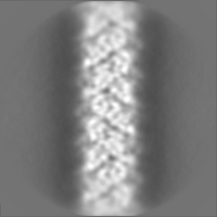
| File | Download / File: emd_6663.map.gz / Format: CCP4 / Size: 30.5 MB / Type: IMAGE STORED AS FLOATING POINT NUMBER (4 BYTES) | ||||||||||||||||||||||||||||||||||||||||||||||||||||||||||||
|---|---|---|---|---|---|---|---|---|---|---|---|---|---|---|---|---|---|---|---|---|---|---|---|---|---|---|---|---|---|---|---|---|---|---|---|---|---|---|---|---|---|---|---|---|---|---|---|---|---|---|---|---|---|---|---|---|---|---|---|---|---|
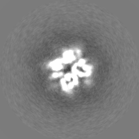
| Projections & slices | Image control
Images are generated by Spider. | ||||||||||||||||||||||||||||||||||||||||||||||||||||||||||||
| Voxel size | X=Y=Z: 1.31 Å | ||||||||||||||||||||||||||||||||||||||||||||||||||||||||||||
| Density |
| ||||||||||||||||||||||||||||||||||||||||||||||||||||||||||||
| Symmetry | Space group: 1 | ||||||||||||||||||||||||||||||||||||||||||||||||||||||||||||
| Details | EMDB XML:
CCP4 map header:
| ||||||||||||||||||||||||||||||||||||||||||||||||||||||||||||
 Z (Sec.)
 Y (Row.)
 X (Col.)


-Supplemental data
-Half map: #1
| File | emd_6663_half_map_1.map | ||||||||||||
|---|---|---|---|---|---|---|---|---|---|---|---|---|---|
| Projections & Slices |
| ||||||||||||
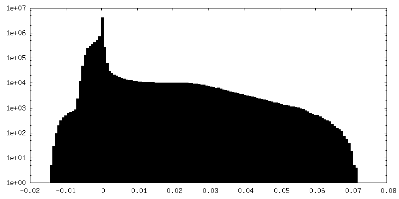
| Density Histograms |


-Half map: #2
| File | emd_6663_half_map_2.map | ||||||||||||
|---|---|---|---|---|---|---|---|---|---|---|---|---|---|
| Projections & Slices |
| ||||||||||||
| Density Histograms |


- Sample components
-Entire : Active RIP2 signaling complex
| Entire | Name: Active RIP2 signaling complex |
|---|---|
| Components |
|
-Supramolecule #1: Active RIP2 signaling complex
| Supramolecule | Name: Active RIP2 signaling complex / type: organelle_or_cellular_component / ID: 1 / Parent: 0 / Macromolecule list: all |
|---|---|
| Source (natural) | Organism: Homo sapiens (human) |
| Recombinant expression | Organism:  |
-Macromolecule #1: Receptor-interacting serine/threonine-protein kinase 2
| Macromolecule | Name: Receptor-interacting serine/threonine-protein kinase 2 type: protein_or_peptide / ID: 1 / Number of copies: 12 / Enantiomer: LEVO / EC number: non-specific serine/threonine protein kinase |
|---|---|
| Source (natural) | Organism: Homo sapiens (human) |
| Molecular weight | Theoretical: 10.751217 KDa |
| Recombinant expression | Organism: |
| Sequence | String: TNSAGIAQQW IQSKREDIVN QMTEACLNQS LDALLSRDLI MKEDYELVST KPTRTSKVRQ LLDTTDIQGE EFAKVIVQKL KDNKQMGLQ PYPEI |
-Experimental details
-Structure determination
| Method | cryo EM |
|---|---|
 Processing Processing | helical reconstruction |
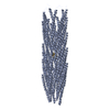
| Aggregation state | filament |
-Sample preparation
| Concentration | 0.2 mg/mL |
|---|---|
| Buffer | pH: 7.4 |
| Grid | Material: COPPER / Support film - Material: CARBON / Support film - topology: HOLEY |
| Vitrification | Cryogen name: METHANE |
- Electron microscopy
| Microscope | FEI TITAN KRIOS |
|---|---|
| Image recording | Film or detector model: FEI FALCON II (4k x 4k) / Average electron dose: 12.6 e/Å2 |
| Electron beam | Acceleration voltage: 200 kV / Electron source:  FIELD EMISSION GUN FIELD EMISSION GUN |
| Electron optics | Illumination mode: FLOOD BEAM / Imaging mode: BRIGHT FIELD |
| Sample stage | Specimen holder model: FEI TITAN KRIOS AUTOGRID HOLDER |
| Experimental equipment |  Model: Titan Krios / Image courtesy: FEI Company |
-Image processing
| Final reconstruction | Applied symmetry - Helical parameters - Δz: 4.9448 Å Applied symmetry - Helical parameters - Δ&Phi: -101.188 ° Applied symmetry - Helical parameters - Axial symmetry: C1 (asymmetric) Resolution.type: BY AUTHOR / Resolution: 4.85 Å / Resolution method: FSC 0.143 CUT-OFF / Software - Name: RELION (ver. 2.0beta) Details: The model is only a partial model. Indefinite filament model could be produced by a helical rotation. Number images used: 118954 |
|---|---|
| CTF correction | Software - Name: RELION (ver. 2.0beta) |
| Final angle assignment | Type: NOT APPLICABLE / Software - Name: RELION (ver. 2.0beta) |
-Atomic model buiding 1
| Refinement | Space: REAL / Protocol: FLEXIBLE FIT |
|---|---|
| Output model |
PDB-5h4a: |
