 Movie
Movie Controller

[English] 日本語
 Yorodumi
- PDB-5x0y: Complex of Snf2-Nucleosome complex with Snf2 bound to SHL2 of the... -
+ Open data

- Basic information
| Entry | Database: PDB / ID: 5x0y | ||||||||||||||||||||||||||||||||||||||||||||||||||||||
|---|---|---|---|---|---|---|---|---|---|---|---|---|---|---|---|---|---|---|---|---|---|---|---|---|---|---|---|---|---|---|---|---|---|---|---|---|---|---|---|---|---|---|---|---|---|---|---|---|---|---|---|---|---|---|---|
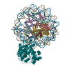
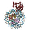
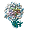
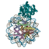
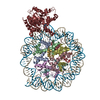
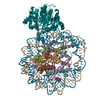
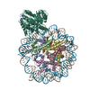
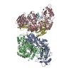
| Title | Complex of Snf2-Nucleosome complex with Snf2 bound to SHL2 of the nucleosome | ||||||||||||||||||||||||||||||||||||||||||||||||||||||
 Components Components |
| ||||||||||||||||||||||||||||||||||||||||||||||||||||||
 Keywords Keywords | STRUCTURAL PROTEIN/HYDROLASE/DNA / Snf2 / nucleosome / chromatin remodeling / STRUCTURAL PROTEIN-HYDROLASE-DNA complex | ||||||||||||||||||||||||||||||||||||||||||||||||||||||
| Function / homology |  Function and homology information Function and homology informationpositive regulation of cell adhesion involved in single-species biofilm formation / positive regulation of mating type switching / positive regulation of invasive growth in response to glucose limitation / aggrephagy / DNA strand invasion / rDNA binding / histone H3K14ac reader activity / nucleosome array spacer activity / ATP-dependent chromatin remodeler activity / SWI/SNF complex ...positive regulation of cell adhesion involved in single-species biofilm formation / positive regulation of mating type switching / positive regulation of invasive growth in response to glucose limitation / aggrephagy / DNA strand invasion / rDNA binding / histone H3K14ac reader activity / nucleosome array spacer activity / ATP-dependent chromatin remodeler activity / SWI/SNF complex / nucleosomal DNA binding / histone H4 reader activity / histone reader activity / transcription initiation-coupled chromatin remodeling / cellular response to amino acid starvation / helicase activity / DNA-templated DNA replication / Hydrolases; Acting on acid anhydrides; Acting on acid anhydrides to facilitate cellular and subcellular movement / structural constituent of chromatin / nucleosome / double-strand break repair / heterochromatin formation / nucleosome assembly / histone binding / RNA polymerase II-specific DNA-binding transcription factor binding / chromatin remodeling / protein heterodimerization activity / hydrolase activity / chromatin binding / regulation of transcription by RNA polymerase II / chromatin / positive regulation of transcription by RNA polymerase II / DNA binding / nucleoplasm / ATP binding / nucleus Similarity search - Function | ||||||||||||||||||||||||||||||||||||||||||||||||||||||
| Biological species |  synthetic construct (others) | ||||||||||||||||||||||||||||||||||||||||||||||||||||||
| Method | ELECTRON MICROSCOPY / single particle reconstruction / cryo EM / Resolution: 4.69 Å | ||||||||||||||||||||||||||||||||||||||||||||||||||||||
 Authors Authors | Li, M. / Liu, X. / Xia, X. / Chen, Z. / Li, X. | ||||||||||||||||||||||||||||||||||||||||||||||||||||||
 Citation Citation | Journal: Nature / Year: 2017 Title: Mechanism of chromatin remodelling revealed by the Snf2-nucleosome structure. Authors: Xiaoyu Liu / Meijing Li / Xian Xia / Xueming Li / Zhucheng Chen /  Abstract: Chromatin remodellers are helicase-like, ATP-dependent enzymes that alter chromatin structure and nucleosome positions to allow regulatory proteins access to DNA. Here we report the cryo-electron ...Chromatin remodellers are helicase-like, ATP-dependent enzymes that alter chromatin structure and nucleosome positions to allow regulatory proteins access to DNA. Here we report the cryo-electron microscopy structure of chromatin remodeller Switch/sucrose non-fermentable (SWI2/SNF2) from Saccharomyces cerevisiae bound to the nucleosome. The structure shows that the two core domains of Snf2 are realigned upon nucleosome binding, suggesting activation of the enzyme. The core domains contact each other through two induced Brace helices, which are crucial for coupling ATP hydrolysis to chromatin remodelling. Snf2 binds to the phosphate backbones of one DNA gyre of the nucleosome mainly through its helicase motifs within the major domain cleft, suggesting a conserved mechanism of substrate engagement across different remodellers. Snf2 contacts the second DNA gyre via a positively charged surface, providing a mechanism to anchor the remodeller at a fixed position of the nucleosome. Snf2 locally deforms nucleosomal DNA at the site of binding, priming the substrate for the remodelling reaction. Together, these findings provide mechanistic insights into chromatin remodelling. | ||||||||||||||||||||||||||||||||||||||||||||||||||||||
| History |
|
- Structure visualization
| Movie |
Movie viewer |
|---|---|
| Structure viewer | Molecule: MolmilJmol/JSmol |
- Downloads & links
-Download
| PDBx/mmCIF format | 5x0y.cif.gz | 440.1 KB | Display | PDBx/mmCIF format |
|---|---|---|---|---|
| PDB format | pdb5x0y.ent.gz | 335.3 KB | Display | PDB format |
| PDBx/mmJSON format | 5x0y.json.gz | Tree view | PDBx/mmJSON format | |
| Others |  Other downloads Other downloads |
-Validation report
| Arichive directory | https://data.pdbj.org/pub/pdb/validation_reports/x0/5x0yftp://data.pdbj.org/pub/pdb/validation_reports/x0/5x0y | HTTPS FTP |
|---|
-Related structure data
| Related structure data | 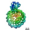 6700MC 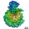 6699C 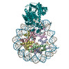 5x0xC M: map data used to model this data C: citing same article ( |
|---|---|
| Similar structure data |
-Links
 PDBj
PDBj


- Assembly
| Deposited unit | 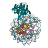
|
|---|---|
| 1 |
|
-Components
-Protein , 5 types, 9 molecules AEBFCGDHO
| #1: Protein | Mass: 15289.904 Da / Num. of mol.: 2 Source method: isolated from a genetically manipulated source Source: (gene. exp.)  #2: Protein | Mass: 11263.231 Da / Num. of mol.: 2 Source method: isolated from a genetically manipulated source Source: (gene. exp.) #3: Protein | Mass: 13978.241 Da / Num. of mol.: 2 Source method: isolated from a genetically manipulated source Source: (gene. exp.) #4: Protein | Mass: 13524.752 Da / Num. of mol.: 2 Source method: isolated from a genetically manipulated source Source: (gene. exp.) #7: Protein | | Mass: 85802.805 Da / Num. of mol.: 1 / Fragment: UNP RESIDUES 666-1400 Source method: isolated from a genetically manipulated source Source: (gene. exp.) Strain: ATCC 204508 / S288c / Gene: SNF2, GAM1, RIC1, SWI2, TYE3, YOR290C / Production host: References: UniProt: P22082, Hydrolases; Acting on acid anhydrides; Acting on acid anhydrides to facilitate cellular and subcellular movement |
|---|
-DNA chain , 2 types, 2 molecules IJ
| #5: DNA chain | Mass: 51381.758 Da / Num. of mol.: 1 / Source method: obtained synthetically / Source: (synth.) synthetic construct (others) |
|---|---|
| #6: DNA chain | Mass: 51723.949 Da / Num. of mol.: 1 / Source method: obtained synthetically / Source: (synth.) synthetic construct (others) |
-Details
| Has protein modification | N |
|---|---|
| Sequence details | Authors state that the sample sequence of chain D/H is conformed by DNA sequencing and consistents ...Authors state that the sample sequence of chain D/H is conformed by DNA sequencing and consistents with the literature (PDB code 3MVD). |
-Experimental details
-Experiment
| Experiment | Method: ELECTRON MICROSCOPY |
|---|---|
| EM experiment | Aggregation state: PARTICLE / 3D reconstruction method: single particle reconstruction |
- Sample preparation
| Component | Name: SHL2 complex / Type: COMPLEX / Entity ID: all / Source: RECOMBINANT |
|---|---|
| Source (natural) | Organism: |
| Source (recombinant) | Organism: |
| Buffer solution | pH: 7 |
| Specimen | Embedding applied: NO / Shadowing applied: NO / Staining applied: NO / Vitrification applied: YES |
| Specimen support | Grid material: COPPER / Grid mesh size: 400 divisions/in. / Grid type: Quantifoil holey carbon grid |
| Vitrification | Instrument: FEI VITROBOT MARK IV / Cryogen name: ETHANE / Humidity: 100 % / Chamber temperature: 281 K |
- Electron microscopy imaging
| Experimental equipment |  Model: Titan Krios / Image courtesy: FEI Company |
|---|---|
| Microscopy | Model: FEI TITAN KRIOS |
| Electron gun | Electron source:  FIELD EMISSION GUN / Accelerating voltage: 300 kV / Illumination mode: FLOOD BEAM FIELD EMISSION GUN / Accelerating voltage: 300 kV / Illumination mode: FLOOD BEAM |
| Electron lens | Mode: BRIGHT FIELD / Nominal magnification: 22500 X / Nominal defocus max: 2500 nm / Nominal defocus min: 1000 nm / Cs: 2.7 mm / C2 aperture diameter: 50 µm / Alignment procedure: COMA FREE |
| Specimen holder | Cryogen: NITROGEN / Specimen holder model: FEI TITAN KRIOS AUTOGRID HOLDER |
| Image recording | Average exposure time: 8 sec. / Electron dose: 50 e/Å2 / Detector mode: SUPER-RESOLUTION / Film or detector model: GATAN K2 SUMMIT (4k x 4k) |
| Image scans | Movie frames/image: 32 |
- Processing
| Software | Name: PHENIX / Version: 1.10.1_2155: / Classification: refinement | ||||||||||||||||||||||||
|---|---|---|---|---|---|---|---|---|---|---|---|---|---|---|---|---|---|---|---|---|---|---|---|---|---|
| EM software |
| ||||||||||||||||||||||||
| CTF correction | Type: PHASE FLIPPING AND AMPLITUDE CORRECTION | ||||||||||||||||||||||||
| 3D reconstruction | Resolution: 4.69 Å / Resolution method: FSC 0.143 CUT-OFF / Num. of particles: 90725 / Symmetry type: POINT | ||||||||||||||||||||||||
| Refine LS restraints |
|
