 Movie
Movie Controller

[English] 日本語
 Yorodumi
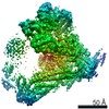
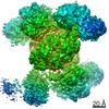
- PDB-5uow: Triheteromeric NMDA receptor GluN1/GluN2A/GluN2B in complex with ... -
+ Open data

- Basic information
| Entry | Database: PDB / ID: 5uow | |||||||||
|---|---|---|---|---|---|---|---|---|---|---|
| Title | Triheteromeric NMDA receptor GluN1/GluN2A/GluN2B in complex with glycine, glutamate, MK-801 and a GluN2B-specific Fab, at pH 6.5 | |||||||||
 Components Components |
| |||||||||
 Keywords Keywords | MEMBRANE PROTEIN | |||||||||
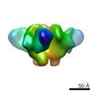
| Function / homology |  Function and homology information Function and homology informationNMDA glutamate receptor activity / NMDA selective glutamate receptor complex / response to zinc ion / ligand-gated sodium channel activity / calcium ion transmembrane import into cytosol / protein heterotetramerization / response to magnesium ion / ligand-gated monoatomic ion channel activity / glutamate-gated calcium ion channel activity / ionotropic glutamate receptor signaling pathway ...NMDA glutamate receptor activity / NMDA selective glutamate receptor complex / response to zinc ion / ligand-gated sodium channel activity / calcium ion transmembrane import into cytosol / protein heterotetramerization / response to magnesium ion / ligand-gated monoatomic ion channel activity / glutamate-gated calcium ion channel activity / ionotropic glutamate receptor signaling pathway / sodium ion transmembrane transport / synaptic transmission, glutamatergic / transmitter-gated monoatomic ion channel activity involved in regulation of postsynaptic membrane potential / regulation of membrane potential / excitatory postsynaptic potential / regulation of synaptic plasticity / postsynaptic density membrane / calcium ion transmembrane transport / long-term synaptic potentiation / late endosome / signaling receptor activity / chemical synaptic transmission / postsynaptic membrane / lysosome / neuron projection / synapse / metal ion binding / plasma membrane Similarity search - Function | |||||||||
| Biological species |  | |||||||||
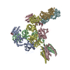
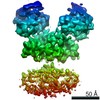
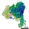
| Method | ELECTRON MICROSCOPY / single particle reconstruction / cryo EM / Resolution: 4.5 Å | |||||||||
 Authors Authors | Lu, W. / Du, J. / Goehring, A. / Gouaux, E. | |||||||||
 Citation Citation | Journal: Science / Year: 2017 Title: Cryo-EM structures of the triheteromeric NMDA receptor and its allosteric modulation. Authors: Wei Lü / Juan Du / April Goehring / Eric Gouaux /  Abstract: -methyl-d-aspartate receptors (NMDARs) are heterotetrameric ion channels assembled as diheteromeric or triheteromeric complexes. Here, we report structures of the triheteromeric GluN1/GluN2A/GluN2B ...-methyl-d-aspartate receptors (NMDARs) are heterotetrameric ion channels assembled as diheteromeric or triheteromeric complexes. Here, we report structures of the triheteromeric GluN1/GluN2A/GluN2B receptor in the absence or presence of the GluN2B-specific allosteric modulator Ro 25-6981 (Ro), determined by cryogenic electron microscopy (cryo-EM). In the absence of Ro, the GluN2A and GluN2B amino-terminal domains (ATDs) adopt "closed" and "open" clefts, respectively. Upon binding Ro, the GluN2B ATD clamshell transitions from an open to a closed conformation. Consistent with a predominance of the GluN2A subunit in ion channel gating, the GluN2A subunit interacts more extensively with GluN1 subunits throughout the receptor, in comparison with the GluN2B subunit. Differences in the conformation of the pseudo-2-fold-related GluN1 subunits further reflect receptor asymmetry. The triheteromeric NMDAR structures provide the first view of the most common NMDA receptor assembly and show how incorporation of two different GluN2 subunits modifies receptor symmetry and subunit interactions, allowing each subunit to uniquely influence receptor structure and function, thus increasing receptor complexity. | |||||||||
| History |
|
- Structure visualization
| Movie |
Movie viewer |
|---|---|
| Structure viewer | Molecule: MolmilJmol/JSmol |
- Downloads & links
-Download
| PDBx/mmCIF format | 5uow.cif.gz | 898 KB | Display | PDBx/mmCIF format |
|---|---|---|---|---|
| PDB format | pdb5uow.ent.gz | 722.1 KB | Display | PDB format |
| PDBx/mmJSON format | 5uow.json.gz | Tree view | PDBx/mmJSON format | |
| Others |  Other downloads Other downloads |
-Validation report
| Arichive directory | https://data.pdbj.org/pub/pdb/validation_reports/uo/5uowftp://data.pdbj.org/pub/pdb/validation_reports/uo/5uow | HTTPS FTP |
|---|
-Related structure data
| Related structure data | 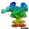 8579MC 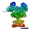 8580C 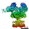 8581C 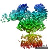 8583C 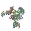 5up2C M: map data used to model this data C: citing same article ( |
|---|---|
| Similar structure data |

-Links
 PDBj
PDBj


- Assembly
| Deposited unit | 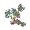
|
|---|---|
| 1 |
|
-Components
-N-methyl-D-aspartate receptor subunit ... , 2 types, 3 molecules ACB
| #1: Protein | Mass: 91639.867 Da / Num. of mol.: 2 Source method: isolated from a genetically manipulated source Source: (gene. exp.)  Homo sapiens (human) / References: UniProt: C0KD18, UniProt: A0A1L8F5J9*PLUS Homo sapiens (human) / References: UniProt: C0KD18, UniProt: A0A1L8F5J9*PLUS#2: Protein | | Mass: 93664.344 Da / Num. of mol.: 1 Source method: isolated from a genetically manipulated source Source: (gene. exp.) Homo sapiens (human) / References: UniProt: B7ZSK1 |
|---|
-Protein / Antibody / Sugars , 3 types, 11 molecules DFG
| #3: Protein | Mass: 94552.234 Da / Num. of mol.: 1 Source method: isolated from a genetically manipulated source Source: (gene. exp.) Homo sapiens (human) / References: UniProt: A7XY94 | ||
|---|---|---|---|
| #4: Antibody | Mass: 18400.619 Da / Num. of mol.: 2 Source method: isolated from a genetically manipulated source Source: (gene. exp.) Homo sapiens (human)#5: Polysaccharide | 2-acetamido-2-deoxy-beta-D-glucopyranose-(1-4)-2-acetamido-2-deoxy-beta-D-glucopyranose Source method: isolated from a genetically manipulated source |
-Non-polymers , 2 types, 2 molecules 
| #6: Chemical | ChemComp-GLU /  Type: L-peptide linking / Mass: 147.129 Da / Num. of mol.: 1 / Source method: obtained synthetically / Formula: C5H9NO4 Type: L-peptide linking / Mass: 147.129 Da / Num. of mol.: 1 / Source method: obtained synthetically / Formula: C5H9NO4 |
|---|---|
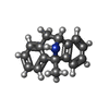
| #7: Chemical | ChemComp-BMK / ( Mass: 221.297 Da / Num. of mol.: 1 / Source method: obtained synthetically / Formula: C16H15N / Comment: neurotransmitter*YM Mass: 221.297 Da / Num. of mol.: 1 / Source method: obtained synthetically / Formula: C16H15N / Comment: neurotransmitter*YM |
-Details
| Has protein modification | Y |
|---|---|
| Sequence details | Sample sequence for this chains is unknown |
-Experimental details
-Experiment
| Experiment | Method: ELECTRON MICROSCOPY |
|---|---|
| EM experiment | Aggregation state: PARTICLE / 3D reconstruction method: single particle reconstruction |
- Sample preparation
| Component | Name: membrane protein / Type: COMPLEX / Entity ID: #1-#4 / Source: RECOMBINANT |
|---|---|
| Source (natural) | Organism: |
| Source (recombinant) | Organism: Homo sapiens (human) |
| Buffer solution | pH: 6.5 |
| Specimen | Embedding applied: NO / Shadowing applied: NO / Staining applied: NO / Vitrification applied: YES |
| Vitrification | Cryogen name: ETHANE |
- Electron microscopy imaging
| Experimental equipment |  Model: Titan Krios / Image courtesy: FEI Company |
|---|---|
| Microscopy | Model: FEI TITAN KRIOS |
| Electron gun | Electron source:  FIELD EMISSION GUN / Accelerating voltage: 300 kV / Illumination mode: FLOOD BEAM FIELD EMISSION GUN / Accelerating voltage: 300 kV / Illumination mode: FLOOD BEAM |
| Electron lens | Mode: BRIGHT FIELD |
| Image recording | Electron dose: 0.84 e/Å2 / Detector mode: SUPER-RESOLUTION / Film or detector model: GATAN K2 SUMMIT (4k x 4k) |
- Processing
| CTF correction | Type: PHASE FLIPPING AND AMPLITUDE CORRECTION |
|---|---|
| 3D reconstruction | Resolution: 4.5 Å / Resolution method: FSC 0.143 CUT-OFF / Num. of particles: 302052 / Symmetry type: POINT |
| Refinement | Highest resolution: 4.5 Å |
