 Movie
Movie Controller


+ Open data

- Basic information
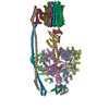
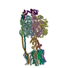
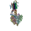
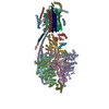
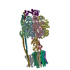
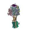
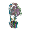
| Entry | Database: PDB / ID: 5t4q | ||||||
|---|---|---|---|---|---|---|---|
| Title | Autoinhibited E. coli ATP synthase state 3 | ||||||
 Components Components | (ATP synthase ...) x 8 | ||||||
 Keywords Keywords | HYDROLASE / ATP synthase / ATPase / rotary motor / membrane protein | ||||||
| Function / homology |  Function and homology information Function and homology informationproton motive force-driven plasma membrane ATP synthesis / proton-transporting two-sector ATPase complex, proton-transporting domain / proton motive force-driven ATP synthesis / proton-transporting ATPase activity, rotational mechanism / H+-transporting two-sector ATPase / proton-transporting ATP synthase complex / proton-transporting ATP synthase activity, rotational mechanism / ADP binding / lipid binding / ATP hydrolysis activity ...proton motive force-driven plasma membrane ATP synthesis / proton-transporting two-sector ATPase complex, proton-transporting domain / proton motive force-driven ATP synthesis / proton-transporting ATPase activity, rotational mechanism / H+-transporting two-sector ATPase / proton-transporting ATP synthase complex / proton-transporting ATP synthase activity, rotational mechanism / ADP binding / lipid binding / ATP hydrolysis activity / ATP binding / membrane / plasma membrane Similarity search - Function | ||||||
| Biological species |  | ||||||
| Method | ELECTRON MICROSCOPY / single particle reconstruction / cryo EM / Resolution: 8.53 Å | ||||||
 Authors Authors | Sobti, M. / Smits, C. / Wong, A.S.W. / Ishmukhametov, R. / Stock, D. / Sandin, S. / Stewart, A.G. | ||||||
 Citation Citation | Journal: Elife / Year: 2016 Title: Cryo-EM structures of the autoinhibited ATP synthase in three rotational states. Authors: Meghna Sobti / Callum Smits / Andrew Sw Wong / Robert Ishmukhametov / Daniela Stock / Sara Sandin / Alastair G Stewart /    Abstract: A molecular model that provides a framework for interpreting the wealth of functional information obtained on the F-ATP synthase has been generated using cryo-electron microscopy. Three different ...A molecular model that provides a framework for interpreting the wealth of functional information obtained on the F-ATP synthase has been generated using cryo-electron microscopy. Three different states that relate to rotation of the enzyme were observed, with the central stalk's ε subunit in an extended autoinhibitory conformation in all three states. The F motor comprises of seven transmembrane helices and a decameric c-ring and invaginations on either side of the membrane indicate the entry and exit channels for protons. The proton translocating subunit contains near parallel helices inclined by ~30° to the membrane, a feature now synonymous with rotary ATPases. For the first time in this rotary ATPase subtype, the peripheral stalk is resolved over its entire length of the complex, revealing the F attachment points and a coiled-coil that bifurcates toward the membrane with its helices separating to embrace subunit from two sides. | ||||||
| History |
|
- Structure visualization
| Movie |
Movie viewer |
|---|---|
| Structure viewer | Molecule: MolmilJmol/JSmol |
- Downloads & links
-Download
| PDBx/mmCIF format | 5t4q.cif.gz | 680.6 KB | Display | PDBx/mmCIF format |
|---|---|---|---|---|
| PDB format | pdb5t4q.ent.gz | 479.1 KB | Display | PDB format |
| PDBx/mmJSON format | 5t4q.json.gz | Tree view | PDBx/mmJSON format | |
| Others |  Other downloads Other downloads |
-Validation report
| Summary document | 5t4q_validation.pdf.gz | 1.2 MB | Display | wwPDB validaton report |
|---|---|---|---|---|
| Full document | 5t4q_full_validation.pdf.gz | 1.2 MB | Display | |
| Data in XML | 5t4q_validation.xml.gz | 98.4 KB | Display | |
| Data in CIF | 5t4q_validation.cif.gz | 164.8 KB | Display | |
| Arichive directory | https://data.pdbj.org/pub/pdb/validation_reports/t4/5t4qftp://data.pdbj.org/pub/pdb/validation_reports/t4/5t4q | HTTPS FTP |
-Related structure data
| Related structure data | 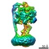 8359MC 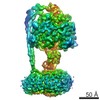 8357C 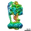 8358C  5t4oC 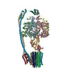 5t4pC M: map data used to model this data C: citing same article ( |
|---|---|
| Similar structure data |


-Links
 PDBj
PDBj


- Assembly
| Deposited unit | 
|
|---|---|
| 1 |
|
-Components
-ATP synthase ... , 8 types, 22 molecules ABCDEFGHIJKLMNOPQRSTUV
| #1: Protein | Mass: 55138.531 Da / Num. of mol.: 3 Source method: isolated from a genetically manipulated source Source: (gene. exp.) References: UniProt: B7MGF4, UniProt: P0ABB0*PLUS, H+-transporting two-sector ATPase #2: Protein | Mass: 51664.574 Da / Num. of mol.: 3 Source method: isolated from a genetically manipulated source Source: (gene. exp.) References: UniProt: B7MGF2, UniProt: P0ABB4*PLUS, H+-transporting two-sector ATPase #3: Protein | | Mass: 31539.285 Da / Num. of mol.: 1 Source method: isolated from a genetically manipulated source Source: (gene. exp.) #4: Protein | | Mass: 15087.244 Da / Num. of mol.: 1 Source method: isolated from a genetically manipulated source Source: (gene. exp.) #5: Protein | Mass: 17126.691 Da / Num. of mol.: 2 Source method: isolated from a genetically manipulated source Source: (gene. exp.) #6: Protein | | Mass: 30324.096 Da / Num. of mol.: 1 Source method: isolated from a genetically manipulated source Source: (gene. exp.) #7: Protein | | Mass: 19289.061 Da / Num. of mol.: 1 Source method: isolated from a genetically manipulated source Source: (gene. exp.) #8: Protein | Mass: 8259.064 Da / Num. of mol.: 10 Source method: isolated from a genetically manipulated source Source: (gene. exp.) |
|---|
-Non-polymers , 2 types, 4 molecules 

| #9: Chemical |  Mass: 507.181 Da / Num. of mol.: 3 / Source method: obtained synthetically / Formula: C10H16N5O13P3 / Comment: ATP, energy-carrying molecule*YM Mass: 507.181 Da / Num. of mol.: 3 / Source method: obtained synthetically / Formula: C10H16N5O13P3 / Comment: ATP, energy-carrying molecule*YM#10: Chemical | ChemComp-ADP / |  Mass: 427.201 Da / Num. of mol.: 1 / Source method: obtained synthetically / Formula: C10H15N5O10P2 / Comment: ADP, energy-carrying molecule*YM Mass: 427.201 Da / Num. of mol.: 1 / Source method: obtained synthetically / Formula: C10H15N5O10P2 / Comment: ADP, energy-carrying molecule*YM |
|---|
-Experimental details
-Experiment
| Experiment | Method: ELECTRON MICROSCOPY |
|---|---|
| EM experiment | Aggregation state: PARTICLE / 3D reconstruction method: single particle reconstruction |
- Sample preparation
| Component | Name: ATP synthase / Type: COMPLEX / Entity ID: all / Source: RECOMBINANT |
|---|---|
| Molecular weight | Value: 0.558 MDa / Experimental value: NO |
| Source (natural) | Organism: |
| Source (recombinant) | Organism: |
| Buffer solution | pH: 8 |
| Specimen | Embedding applied: NO / Shadowing applied: NO / Staining applied: NO / Vitrification applied: YES |
| Vitrification | Cryogen name: ETHANE |
- Electron microscopy imaging
| Experimental equipment |  Model: Titan Krios / Image courtesy: FEI Company |
|---|---|
| Microscopy | Model: FEI TITAN KRIOS |
| Electron gun | Electron source:  FIELD EMISSION GUN / Accelerating voltage: 300 kV / Illumination mode: FLOOD BEAM FIELD EMISSION GUN / Accelerating voltage: 300 kV / Illumination mode: FLOOD BEAM |
| Electron lens | Mode: BRIGHT FIELD |
| Image recording | Electron dose: 29 e/Å2 / Film or detector model: FEI FALCON II (4k x 4k) |
- Processing
| CTF correction | Type: PHASE FLIPPING AND AMPLITUDE CORRECTION |
|---|---|
| 3D reconstruction | Resolution: 8.53 Å / Resolution method: FSC 0.143 CUT-OFF / Num. of particles: 95345 / Symmetry type: POINT |
