[English] 日本語
 Yorodumi
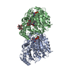
- PDB-5sye: Near-atomic resolution cryo-EM reconstruction of doubly bound Tax... -
+ Open data
- Basic information
| Entry | Database: PDB / ID: 5sye | ||||||||||||
|---|---|---|---|---|---|---|---|---|---|---|---|---|---|
| Title | Near-atomic resolution cryo-EM reconstruction of doubly bound Taxol- and peloruside-stabilized microtubule | ||||||||||||
 Components Components |
| ||||||||||||
 Keywords Keywords | STRUCTURAL PROTEIN / Taxol / peloruside / microtubule | ||||||||||||
| Function / homology |  Function and homology information Function and homology informationMicrotubule-dependent trafficking of connexons from Golgi to the plasma membrane / Resolution of Sister Chromatid Cohesion / Hedgehog 'off' state / Cilium Assembly / Intraflagellar transport / COPI-dependent Golgi-to-ER retrograde traffic / Mitotic Prometaphase / Carboxyterminal post-translational modifications of tubulin / RHOH GTPase cycle / EML4 and NUDC in mitotic spindle formation ...Microtubule-dependent trafficking of connexons from Golgi to the plasma membrane / Resolution of Sister Chromatid Cohesion / Hedgehog 'off' state / Cilium Assembly / Intraflagellar transport / COPI-dependent Golgi-to-ER retrograde traffic / Mitotic Prometaphase / Carboxyterminal post-translational modifications of tubulin / RHOH GTPase cycle / EML4 and NUDC in mitotic spindle formation / Sealing of the nuclear envelope (NE) by ESCRT-III / Kinesins / PKR-mediated signaling / Separation of Sister Chromatids / The role of GTSE1 in G2/M progression after G2 checkpoint / Aggrephagy / RHO GTPases activate IQGAPs / RHO GTPases Activate Formins / HSP90 chaperone cycle for steroid hormone receptors (SHR) in the presence of ligand / MHC class II antigen presentation / Recruitment of NuMA to mitotic centrosomes / COPI-mediated anterograde transport / microtubule-based process / structural constituent of cytoskeleton / microtubule cytoskeleton organization / neuron migration / mitotic cell cycle / microtubule cytoskeleton / Hydrolases; Acting on acid anhydrides; Acting on GTP to facilitate cellular and subcellular movement / microtubule / hydrolase activity / GTPase activity / GTP binding / metal ion binding / cytoplasm Similarity search - Function | ||||||||||||
| Biological species |  | ||||||||||||
| Method | ELECTRON MICROSCOPY / helical reconstruction / cryo EM / Resolution: 3.5 Å | ||||||||||||
 Authors Authors | Kellogg, E.H. / Nogales, E. | ||||||||||||
| Funding support |  United States, United States,  Spain, 3items Spain, 3items
| ||||||||||||
 Citation Citation |  Journal: J Mol Biol / Year: 2017 Journal: J Mol Biol / Year: 2017Title: Insights into the Distinct Mechanisms of Action of Taxane and Non-Taxane Microtubule Stabilizers from Cryo-EM Structures. Authors: Elizabeth H Kellogg / Nisreen M A Hejab / Stuart Howes / Peter Northcote / John H Miller / J Fernando Díaz / Kenneth H Downing / Eva Nogales /    Abstract: A number of microtubule (MT)-stabilizing agents (MSAs) have demonstrated or predicted potential as anticancer agents, but a detailed structural basis for their mechanism of action is still lacking. ...A number of microtubule (MT)-stabilizing agents (MSAs) have demonstrated or predicted potential as anticancer agents, but a detailed structural basis for their mechanism of action is still lacking. We have obtained high-resolution (3.9-4.2Å) cryo-electron microscopy (cryo-EM) reconstructions of MTs stabilized by the taxane-site binders Taxol and zampanolide, and by peloruside, which targets a distinct, non-taxoid pocket on β-tubulin. We find that each molecule has unique distinct structural effects on the MT lattice structure. Peloruside acts primarily at lateral contacts and has an effect on the "seam" of heterologous interactions, enforcing a conformation more similar to that of homologous (i.e., non-seam) contacts by which it regularizes the MT lattice. In contrast, binding of either Taxol or zampanolide induces MT heterogeneity. In doubly bound MTs, peloruside overrides the heterogeneity induced by Taxol binding. Our structural analysis illustrates distinct mechanisms of these drugs for stabilizing the MT lattice and is of relevance to the possible use of combinations of MSAs to regulate MT activity and improve therapeutic potential. | ||||||||||||
| History |
|
- Structure visualization
| Movie |
 Movie viewer Movie viewer |
|---|---|
| Structure viewer | Molecule:  Molmil Molmil Jmol/JSmol Jmol/JSmol |
- Downloads & links
- Download
| PDBx/mmCIF format |  5sye.cif.gz 5sye.cif.gz | 185.3 KB | Display |  PDBx/mmCIF format PDBx/mmCIF format |
|---|---|---|---|---|
| PDB format |  pdb5sye.ent.gz pdb5sye.ent.gz | 142.8 KB | Display |  PDB format PDB format |
| PDBx/mmJSON format |  5sye.json.gz 5sye.json.gz | Tree view |  PDBx/mmJSON format PDBx/mmJSON format | |
| Others |  Other downloads Other downloads |
-Validation report
| Summary document |  5sye_validation.pdf.gz 5sye_validation.pdf.gz | 1.3 MB | Display |  wwPDB validaton report wwPDB validaton report |
|---|---|---|---|---|
| Full document |  5sye_full_validation.pdf.gz 5sye_full_validation.pdf.gz | 1.3 MB | Display | |
| Data in XML |  5sye_validation.xml.gz 5sye_validation.xml.gz | 35.3 KB | Display | |
| Data in CIF |  5sye_validation.cif.gz 5sye_validation.cif.gz | 49.6 KB | Display | |
| Arichive directory |  https://data.pdbj.org/pub/pdb/validation_reports/sy/5sye https://data.pdbj.org/pub/pdb/validation_reports/sy/5sye ftp://data.pdbj.org/pub/pdb/validation_reports/sy/5sye ftp://data.pdbj.org/pub/pdb/validation_reports/sy/5sye | HTTPS FTP |
-Related structure data
| Related structure data |  8321MC  8320C  8322C  8323C  5sycC  5syfC  5sygC M: map data used to model this data C: citing same article ( |
|---|---|
| Similar structure data |
- Links
Links
- Assembly
| Deposited unit | 
|
|---|---|
| 1 | x 26
|
| 2 |
|
| 3 | 
|
| Symmetry | Helical symmetry: (Circular symmetry: 1 / Dyad axis: no / N subunits divisor: 1 / Num. of operations: 26 / Rise per n subunits: 9.43 Å / Rotation per n subunits: -27.7 °) |
- Components
-Protein , 2 types, 2 molecules AB
| #1: Protein | Mass: 48679.051 Da / Num. of mol.: 1 / Source method: isolated from a natural source / Source: (natural)  |
|---|---|
| #2: Protein | Mass: 47825.859 Da / Num. of mol.: 1 / Source method: isolated from a natural source / Source: (natural)  |
-Non-polymers , 5 types, 5 molecules 


| #3: Chemical | ChemComp-GTP / |
|---|---|
| #4: Chemical | ChemComp-MG / |
| #5: Chemical | ChemComp-GDP / |
| #6: Chemical | ChemComp-TA1 / |
| #7: Chemical | ChemComp-POU / |
-Experimental details
-Experiment
| Experiment | Method: ELECTRON MICROSCOPY |
|---|---|
| EM experiment | Aggregation state: HELICAL ARRAY / 3D reconstruction method: helical reconstruction |
- Sample preparation
| Component | Name: Ternary complex of Taxol- and peloruside-stabilized microtubule lattice, alpha-tubulin, and beta-tubulin Type: COMPLEX / Entity ID: #1-#2 / Source: NATURAL | ||||||||||||||||||||||||||||||
|---|---|---|---|---|---|---|---|---|---|---|---|---|---|---|---|---|---|---|---|---|---|---|---|---|---|---|---|---|---|---|---|
| Molecular weight | Experimental value: NO | ||||||||||||||||||||||||||||||
| Source (natural) | Organism:  | ||||||||||||||||||||||||||||||
| Buffer solution | pH: 6.8 | ||||||||||||||||||||||||||||||
| Buffer component |
| ||||||||||||||||||||||||||||||
| Specimen | Conc.: 0.4 mg/ml / Embedding applied: NO / Shadowing applied: NO / Staining applied: NO / Vitrification applied: YES | ||||||||||||||||||||||||||||||
| Specimen support | Grid material: COPPER / Grid mesh size: 400 divisions/in. / Grid type: Protochips CF-1.2/1.3 | ||||||||||||||||||||||||||||||
| Vitrification | Instrument: FEI VITROBOT MARK IV / Cryogen name: ETHANE / Humidity: 100 % / Chamber temperature: 277.15 K Details: Blot for 4 seconds, blot force 10, before plunging into liquid ethane (FEI VITROBOT MARK IV). |
- Electron microscopy imaging
| Microscopy | Model: FEI TITAN |
|---|---|
| Electron gun | Electron source:  FIELD EMISSION GUN / Accelerating voltage: 300 kV / Illumination mode: FLOOD BEAM FIELD EMISSION GUN / Accelerating voltage: 300 kV / Illumination mode: FLOOD BEAM |
| Electron lens | Mode: BRIGHT FIELD / Nominal magnification: 27500 X / Nominal defocus max: 2500 nm / Nominal defocus min: 1000 nm / Calibrated defocus min: 1100 nm / Calibrated defocus max: 3800 nm / Cs: 2.7 mm / C2 aperture diameter: 50 µm / Alignment procedure: COMA FREE |
| Specimen holder | Cryogen: NITROGEN Specimen holder model: GATAN 626 SINGLE TILT LIQUID NITROGEN CRYO TRANSFER HOLDER Temperature (max): 93 K / Temperature (min): 93 K |
| Image recording | Average exposure time: 0.3 sec. / Electron dose: 8 e/Å2 / Detector mode: COUNTING / Film or detector model: GATAN K2 SUMMIT (4k x 4k) / Num. of grids imaged: 1 / Num. of real images: 312 |
| Image scans | Sampling size: 5 µm / Width: 3710 / Height: 3838 / Movie frames/image: 20 / Used frames/image: 1-20 |
- Processing
| EM software |
| ||||||||||||||||||||||||||||||||||||||||||||||||||
|---|---|---|---|---|---|---|---|---|---|---|---|---|---|---|---|---|---|---|---|---|---|---|---|---|---|---|---|---|---|---|---|---|---|---|---|---|---|---|---|---|---|---|---|---|---|---|---|---|---|---|---|
| CTF correction | Type: PHASE FLIPPING AND AMPLITUDE CORRECTION | ||||||||||||||||||||||||||||||||||||||||||||||||||
| Helical symmerty | Angular rotation/subunit: -27.7 ° / Axial rise/subunit: 9.43 Å / Axial symmetry: C1 | ||||||||||||||||||||||||||||||||||||||||||||||||||
| Particle selection | Num. of particles selected: 27275 Details: Microtubule filaments were manually selected using manualpicker.py. | ||||||||||||||||||||||||||||||||||||||||||||||||||
| 3D reconstruction | Resolution: 3.5 Å / Resolution method: FSC 0.143 CUT-OFF / Num. of particles: 17069 / Algorithm: FOURIER SPACE / Symmetry type: HELICAL | ||||||||||||||||||||||||||||||||||||||||||||||||||
| Atomic model building | Protocol: FLEXIBLE FIT / Space: REAL / Target criteria: correlation coefficient |
 Movie
Movie Controller


 PDBj
PDBj


