 Movie
Movie Controller

[English] 日本語
 Yorodumi
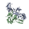
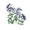
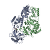
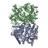
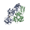
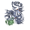
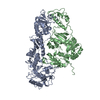
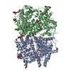
- PDB-5nl2: cryo-EM structure of the mTMEM16A ion channel at 6.6 A resolution. -
+ Open data

- Basic information
| Entry | Database: PDB / ID: 5nl2 | |||||||||
|---|---|---|---|---|---|---|---|---|---|---|
| Title | cryo-EM structure of the mTMEM16A ion channel at 6.6 A resolution. | |||||||||
 Components Components | Anoctamin-1 | |||||||||
 Keywords Keywords | MEMBRANE PROTEIN / TMEM16 family / ion channel / cryo-EM | |||||||||
| Function / homology |  Function and homology information Function and homology informationglial cell projection elongation / trachea development / iodide transmembrane transporter activity / mucus secretion / iodide transport / intracellularly calcium-gated chloride channel activity / cellular response to peptide / Stimuli-sensing channels / voltage-gated chloride channel activity / calcium-activated cation channel activity ...glial cell projection elongation / trachea development / iodide transmembrane transporter activity / mucus secretion / iodide transport / intracellularly calcium-gated chloride channel activity / cellular response to peptide / Stimuli-sensing channels / voltage-gated chloride channel activity / calcium-activated cation channel activity / protein localization to membrane / chloride transport / chloride channel activity / detection of temperature stimulus involved in sensory perception of pain / chloride channel complex / chloride transmembrane transport / regulation of membrane potential / cellular response to heat / presynaptic membrane / phospholipase C-activating G protein-coupled receptor signaling pathway / apical plasma membrane / external side of plasma membrane / signaling receptor binding / glutamatergic synapse / protein homodimerization activity / nucleoplasm / metal ion binding / identical protein binding / plasma membrane Similarity search - Function | |||||||||
| Biological species |  | |||||||||
| Method | ELECTRON MICROSCOPY / single particle reconstruction / cryo EM / Resolution: 6.6 Å | |||||||||
 Authors Authors | Paulino, C. / Neldner, Y. / Lam, K.M. / Kalienkova, V. / Brunner, J.D. / Schenck, S. / Dutzler, R. | |||||||||
| Funding support |  Switzerland, 2items Switzerland, 2items
| |||||||||
 Citation Citation | Journal: Elife / Year: 2017 Title: Structural basis for anion conduction in the calcium-activated chloride channel TMEM16A. Authors: Cristina Paulino / Yvonne Neldner / Andy Km Lam / Valeria Kalienkova / Janine Denise Brunner / Stephan Schenck / Raimund Dutzler / Abstract: The calcium-activated chloride channel TMEM16A is a member of a conserved protein family that comprises ion channels and lipid scramblases. Although the structure of the scramblase nhTMEM16 has ...The calcium-activated chloride channel TMEM16A is a member of a conserved protein family that comprises ion channels and lipid scramblases. Although the structure of the scramblase nhTMEM16 has defined the architecture of the family, it was unknown how a channel has adapted to cope with its distinct functional properties. Here we have addressed this question by the structure determination of mouse TMEM16A by cryo-electron microscopy and a complementary functional characterization. The protein shows a similar organization to nhTMEM16, except for changes at the site of catalysis. There, the conformation of transmembrane helices constituting a membrane-spanning furrow that provides a path for lipids in scramblases has changed to form an enclosed aqueous pore that is largely shielded from the membrane. Our study thus reveals the structural basis of anion conduction in a TMEM16 channel and it defines the foundation for the diverse functional behavior in the TMEM16 family. | |||||||||
| History |
|
- Structure visualization
| Movie |
Movie viewer |
|---|---|
| Structure viewer | Molecule: MolmilJmol/JSmol |
- Downloads & links
-Download
| PDBx/mmCIF format | 5nl2.cif.gz | 151.3 KB | Display | PDBx/mmCIF format |
|---|---|---|---|---|
| PDB format | pdb5nl2.ent.gz | 91.6 KB | Display | PDB format |
| PDBx/mmJSON format | 5nl2.json.gz | Tree view | PDBx/mmJSON format | |
| Others |  Other downloads Other downloads |
-Validation report
| Arichive directory | https://data.pdbj.org/pub/pdb/validation_reports/nl/5nl2ftp://data.pdbj.org/pub/pdb/validation_reports/nl/5nl2 | HTTPS FTP |
|---|
-Related structure data
| Related structure data |  3658MC M: map data used to model this data C: citing same article ( |
|---|---|
| Similar structure data |


-Links
 PDBj
PDBj- Assembly
| Deposited unit | 
|
|---|---|
| 1 |
|
-Components
| #1: Protein | Mass: 111058.992 Da / Num. of mol.: 2 Source method: isolated from a genetically manipulated source Source: (gene. exp.) Cell line (production host): HEK293 stable mTMEM16A cell line (Flp-In System) Production host:  Homo sapiens (human) / References: UniProt: Q8BHY3 Homo sapiens (human) / References: UniProt: Q8BHY3 |
|---|
-Experimental details
-Experiment
| Experiment | Method: ELECTRON MICROSCOPY |
|---|---|
| EM experiment | Aggregation state: PARTICLE / 3D reconstruction method: single particle reconstruction |
- Sample preparation
| Component | Name: mTMEM16A / Type: COMPLEX / Entity ID: all / Source: RECOMBINANT |
|---|---|
| Molecular weight | Value: 0.110916 MDa / Experimental value: YES |
| Source (natural) | Organism: |
| Source (recombinant) | Organism: Homo sapiens (human) / Strain: HEK293 / Cell: stabel mTMEM16A cell line (Flp-In System) |
| Buffer solution | pH: 7.5 / Details: 20 mM Hepes 150 mM NaCl 0.5 mM CaCl2 <0.12% digitonin |
| Buffer component | Conc.: 20 mM / Name: Hepes |
| Specimen | Conc.: 3.08 mg/ml / Embedding applied: NO / Shadowing applied: NO / Staining applied: NO / Vitrification applied: YES Details: full-length (wild-type isoform ac) deglycosylated mTMEM16A |
| Specimen support | Grid material: GOLD / Grid mesh size: 200 divisions/in. / Grid type: Quantifoil R1.2/1.3 |
| Vitrification | Instrument: FEI VITROBOT MARK IV / Cryogen name: ETHANE / Humidity: 100 % / Chamber temperature: 293 K / Details: 2.5 ul sample volume 1-5 sec blotting time |
- Electron microscopy imaging
| Experimental equipment |  Model: Titan Krios / Image courtesy: FEI Company |
|---|---|
| Microscopy | Model: FEI TITAN KRIOS |
| Electron gun | Electron source:  FIELD EMISSION GUN / Accelerating voltage: 300 kV / Illumination mode: FLOOD BEAM FIELD EMISSION GUN / Accelerating voltage: 300 kV / Illumination mode: FLOOD BEAM |
| Electron lens | Mode: BRIGHT FIELD / Nominal magnification: 37037 X / Calibrated magnification: 37037 X / Nominal defocus max: 3800 nm / Nominal defocus min: 500 nm / Calibrated defocus min: 500 nm / Calibrated defocus max: 3800 nm / Cs: 2.7 mm / C2 aperture diameter: 100 µm / Alignment procedure: COMA FREE |
| Specimen holder | Cryogen: NITROGEN / Specimen holder model: FEI TITAN KRIOS AUTOGRID HOLDER / Temperature (max): 100 K / Temperature (min): 80 K |
| Image recording | Average exposure time: 15 sec. / Electron dose: 80 e/Å2 / Detector mode: SUPER-RESOLUTION / Film or detector model: GATAN K2 SUMMIT (4k x 4k) / Num. of grids imaged: 5 / Num. of real images: 5503 Details: Data were collected in an automated fashion on a K2 Summit detector (Gatan) set to super-resolution mode with a pixel size of 0.675 A and a defocus range of -0.5 to -3.8 um using SerialEM. ...Details: Data were collected in an automated fashion on a K2 Summit detector (Gatan) set to super-resolution mode with a pixel size of 0.675 A and a defocus range of -0.5 to -3.8 um using SerialEM. Images were recorded for 15 sec with an initial sub-frame exposure time of 300 ms (50 frames total) with a dose of 1.5 e-/A^2/frame, and later with a sub-frame exposure time of 150 ms (100 frames total) with a dose of 0.8 e-/A^2/frame, resulting in a total accumulated dose on the specimen level of approximately 80 e-/A^2. |
| EM imaging optics | Energyfilter name: GIF / Energyfilter upper: 10 eV / Energyfilter lower: -10 eV |
| Image scans | Sampling size: 5 µm / Width: 7420 / Height: 7676 / Movie frames/image: 50 / Used frames/image: 1-50 |
- Processing
| Software | Name: PHENIX / Version: 1.11.1_2575: / Classification: refinement | ||||||||||||||||||||||||||||||||||||||||
|---|---|---|---|---|---|---|---|---|---|---|---|---|---|---|---|---|---|---|---|---|---|---|---|---|---|---|---|---|---|---|---|---|---|---|---|---|---|---|---|---|---|
| EM software |
| ||||||||||||||||||||||||||||||||||||||||
| Image processing | Details: Fourier cropping (final pixel size 1.35 A), motion correction and dose-weighting of frames were performed by MotionCor2 | ||||||||||||||||||||||||||||||||||||||||
| CTF correction | Details: The contrast transfer function (CTF) parameters were estimated on the movie frames by ctffind4.1 Type: PHASE FLIPPING AND AMPLITUDE CORRECTION | ||||||||||||||||||||||||||||||||||||||||
| Particle selection | Num. of particles selected: 755348 Details: Images showing a strong drift, higher defocus than -3.8 um or a bad CTF estimation were discarded, resulting in 4,178 images used for further analysis. Image processing was performed using ...Details: Images showing a strong drift, higher defocus than -3.8 um or a bad CTF estimation were discarded, resulting in 4,178 images used for further analysis. Image processing was performed using the software package RELION1.4 and at a later stage RELION2.0. Approximately 4,000 particles were manually picked to generate templates for automated particle selection. Following automated picking in RELION, false positives were eliminated manually or through a first round of 2D classification resulting in 755,348 particles. | ||||||||||||||||||||||||||||||||||||||||
| Symmetry | Point symmetry: C2 (2 fold cyclic) | ||||||||||||||||||||||||||||||||||||||||
| 3D reconstruction | Resolution: 6.6 Å / Resolution method: FSC 0.143 CUT-OFF / Num. of particles: 213243 / Symmetry type: POINT | ||||||||||||||||||||||||||||||||||||||||
| Atomic model building | Protocol: RIGID BODY FIT | ||||||||||||||||||||||||||||||||||||||||
| Atomic model building | PDB-ID: 4WIT Accession code: 4WIT / Source name: PDB / Type: experimental model | ||||||||||||||||||||||||||||||||||||||||
| Refine LS restraints |
|

