[English] 日本語
Yorodumi
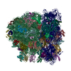
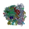
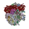
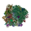
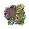
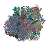
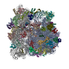
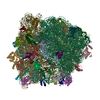
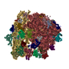
- PDB-5iqr: Structure of RelA bound to the 70S ribosome -

+
Open data


ID or keywords:

Loading...

-
Basic information

Entry
Database: PDB / ID: 5iqr
TitleStructure of RelA bound to the 70S ribosome
Components
  • (30S ribosomal protein ...) x 20
  • (50S ribosomal protein ...) x 31
  • 5S rRNA
  • A/T tRNA(Phe)
  • E-site tRNA(Phe)
  • GTP pyrophosphokinase
  • LSU rRNA
  • P-site fMet-tRNA(fMet)
  • SSU rRNA
  • mRNA
KeywordsRIBOSOME / tRNA / RelA / ppGpp
Function / homology
Function and homology information


guanosine tetraphosphate metabolic process / guanosine-3',5'-bis(diphosphate) 3'-diphosphatase activity / GTP diphosphokinase / GTP diphosphokinase activity / guanosine tetraphosphate biosynthetic process / nucleobase-containing small molecule interconversion / negative regulation of cytoplasmic translational initiation / response to starvation / stringent response / ornithine decarboxylase inhibitor activity ...guanosine tetraphosphate metabolic process / guanosine-3',5'-bis(diphosphate) 3'-diphosphatase activity / GTP diphosphokinase / GTP diphosphokinase activity / guanosine tetraphosphate biosynthetic process / nucleobase-containing small molecule interconversion / negative regulation of cytoplasmic translational initiation / response to starvation / stringent response / ornithine decarboxylase inhibitor activity / transcription antitermination factor activity, RNA binding / misfolded RNA binding / Group I intron splicing / RNA folding / transcriptional attenuation / endoribonuclease inhibitor activity / RNA-binding transcription regulator activity / positive regulation of ribosome biogenesis / negative regulation of cytoplasmic translation / translational termination / four-way junction DNA binding / DnaA-L2 complex / translation repressor activity / negative regulation of DNA-templated DNA replication initiation / negative regulation of translational initiation / regulation of mRNA stability / mRNA regulatory element binding translation repressor activity / ribosome assembly / assembly of large subunit precursor of preribosome / positive regulation of RNA splicing / transcription elongation factor complex / cytosolic ribosome assembly / regulation of DNA-templated transcription elongation / DNA endonuclease activity / response to reactive oxygen species / transcription antitermination / regulation of cell growth / translational initiation / DNA-templated transcription termination / maintenance of translational fidelity / response to radiation / mRNA 5'-UTR binding / ribosomal small subunit biogenesis / small ribosomal subunit rRNA binding / large ribosomal subunit / ribosome biogenesis / ribosome binding / kinase activity / regulation of translation / ribosomal small subunit assembly / small ribosomal subunit / 5S rRNA binding / large ribosomal subunit rRNA binding / transferase activity / cytosolic small ribosomal subunit / ribosomal large subunit assembly / cytoplasmic translation / cytosolic large ribosomal subunit / tRNA binding / molecular adaptor activity / negative regulation of translation / rRNA binding / ribosome / structural constituent of ribosome / translation / response to antibiotic / negative regulation of DNA-templated transcription / mRNA binding / GTP binding / DNA binding / RNA binding / zinc ion binding / ATP binding / membrane / cytosol / cytoplasm
Similarity search - Function
RelA/SpoT, AH and RIS domains / RelA/SpoT, AH and RIS domains / RelA/SpoT family / RelA/SpoT, TGS domain / ACT domain / HD domain / Region found in RelA / SpoT proteins / RelA/SpoT / Region found in RelA / SpoT proteins / TGS domain ...RelA/SpoT, AH and RIS domains / RelA/SpoT, AH and RIS domains / RelA/SpoT family / RelA/SpoT, TGS domain / ACT domain / HD domain / Region found in RelA / SpoT proteins / RelA/SpoT / Region found in RelA / SpoT proteins / TGS domain / ACT domain profile. / ACT domain / HD domain profile. / ACT-like domain / TGS-like / TGS domain profile. / TGS / HD domain / Ribosomal protein L10, eubacterial, conserved site / Ribosomal protein L10 signature. / Ribosomal protein L10 / : / Beta-grasp domain superfamily / Ribosomal protein S21, conserved site / Ribosomal protein S21 signature. / Ribosomal protein L25, short-form / Ribosomal protein S14, bacterial/plastid / Ribosomal protein L11, bacterial-type / Ribosomal protein L31 type A / Ribosomal protein S21 superfamily / Ribosomal protein S21 / Ribosomal protein S16, conserved site / Ribosomal protein S16 signature. / Ribosomal protein S21 / Ribosomal protein L31 signature. / Ribosomal protein L31 / Ribosomal protein L31 superfamily / Ribosomal protein L31 / Nucleotidyltransferase superfamily / Ribosomal protein L11, conserved site / : / Ribosomal protein L11 signature. / Ribosomal protein L21, conserved site / Ribosomal protein L21 signature. / Ribosomal protein L10-like domain superfamily / Ribosomal protein L10P / Ribosomal protein L10 / Ribosomal protein L16 signature 1. / : / Ribosomal protein L6, conserved site / Ribosomal protein L6 signature 1. / Ribosomal protein L16, conserved site / Ribosomal protein L16 signature 2. / Ribosomal protein L9 signature. / Ribosomal protein L9, bacteria/chloroplast / Ribosomal protein L9, C-terminal / Ribosomal protein L9, C-terminal domain / Ribosomal protein L9, C-terminal domain superfamily / Ribosomal protein L17 signature. / Ribosomal protein L11, N-terminal / Ribosomal L25p family / Ribosomal protein L11, N-terminal domain / Ribosomal protein L25 / Ribosomal protein L11/L12 / Ribosomal protein L11, C-terminal / Ribosomal protein L11, C-terminal domain superfamily / Ribosomal protein L11/L12, N-terminal domain superfamily / Ribosomal protein L11, RNA binding domain / Ribosomal protein L11/L12 / Ribosomal protein L36 signature. / Ribosomal protein L28/L24 superfamily / Ribosomal protein L25/Gln-tRNA synthetase, N-terminal / Ribosomal protein L25/Gln-tRNA synthetase, anti-codon-binding domain superfamily / Ribosomal protein L32p, bacterial type / Ribosomal protein L9, N-terminal domain superfamily / Ribosomal protein L9 / Ribosomal protein L9, N-terminal / Ribosomal protein L9, N-terminal domain / : / : / Ribosomal protein L28 / Ribosomal protein L35, conserved site / Ribosomal protein L35 signature. / Ribosomal protein L33, conserved site / Ribosomal protein L33 signature. / Ribosomal protein L35, non-mitochondrial / Ribosomal protein L5, bacterial-type / Ribosomal protein L18, bacterial-type / Ribosomal protein L6, bacterial-type / Ribosomal protein L9/RNase H1, N-terminal / Ribosomal protein L19, conserved site / Ribosomal protein L19 signature. / Ribosomal protein S3, bacterial-type / Ribosomal protein S6, conserved site / Ribosomal protein S6 signature. / Ribosomal protein S19, bacterial-type / Ribosomal protein S13, bacterial-type / Ribosomal protein L36 / Ribosomal protein L36 superfamily / Ribosomal protein L36
Similarity search - Domain/homology
METHIONINE / PAROMOMYCIN / RNA / RNA (> 10) / RNA (> 100) / RNA (> 1000) / Small ribosomal subunit protein bS6 / Small ribosomal subunit protein uS7 / Large ribosomal subunit protein uL15 / Large ribosomal subunit protein uL10 ...METHIONINE / PAROMOMYCIN / RNA / RNA (> 10) / RNA (> 100) / RNA (> 1000) / Small ribosomal subunit protein bS6 / Small ribosomal subunit protein uS7 / Large ribosomal subunit protein uL15 / Large ribosomal subunit protein uL10 / Large ribosomal subunit protein uL11 / Large ribosomal subunit protein bL19 / Large ribosomal subunit protein bL20 / Large ribosomal subunit protein bL27 / Large ribosomal subunit protein bL28 / Large ribosomal subunit protein uL29 / Large ribosomal subunit protein bL31 / Large ribosomal subunit protein bL32 / Large ribosomal subunit protein bL33 / Large ribosomal subunit protein bL34 / Large ribosomal subunit protein bL35 / Large ribosomal subunit protein bL36A / Large ribosomal subunit protein bL9 / Small ribosomal subunit protein uS10 / Small ribosomal subunit protein uS11 / Small ribosomal subunit protein uS12 / Small ribosomal subunit protein uS13 / Small ribosomal subunit protein bS16 / Small ribosomal subunit protein bS18 / Small ribosomal subunit protein uS19 / Small ribosomal subunit protein bS20 / Small ribosomal subunit protein uS2 / Small ribosomal subunit protein uS3 / Small ribosomal subunit protein uS4 / Small ribosomal subunit protein uS5 / Small ribosomal subunit protein uS8 / Small ribosomal subunit protein uS9 / Large ribosomal subunit protein uL13 / Large ribosomal subunit protein uL14 / Large ribosomal subunit protein uL16 / Large ribosomal subunit protein uL23 / Small ribosomal subunit protein uS15 / GTP pyrophosphokinase / Large ribosomal subunit protein bL17 / Large ribosomal subunit protein bL21 / Large ribosomal subunit protein uL30 / Large ribosomal subunit protein uL6 / Small ribosomal subunit protein uS14 / Small ribosomal subunit protein uS17 / Large ribosomal subunit protein uL18 / Large ribosomal subunit protein uL2 / Large ribosomal subunit protein uL3 / Large ribosomal subunit protein uL24 / Large ribosomal subunit protein uL4 / Large ribosomal subunit protein uL22 / Large ribosomal subunit protein uL5 / Small ribosomal subunit protein bS21 / Large ribosomal subunit protein bL25
Similarity search - Component
Biological speciesEscherichia coli K-12 (bacteria)
synthetic construct (others)
MethodELECTRON MICROSCOPY / single particle reconstruction / cryo EM / Resolution: 3 Å
AuthorsBrown, A. / Fernandez, I.S. / Gordiyenko, Y. / Ramakrishnan, V.
CitationJournal: Nature / Year: 2016
Title: Ribosome-dependent activation of stringent control.
Authors: Alan Brown / Israel S Fernández / Yuliya Gordiyenko / V Ramakrishnan /
Abstract: In order to survive, bacteria continually sense, and respond to, environmental fluctuations. Stringent control represents a key bacterial stress response to nutrient starvation that leads to rapid ...In order to survive, bacteria continually sense, and respond to, environmental fluctuations. Stringent control represents a key bacterial stress response to nutrient starvation that leads to rapid and comprehensive reprogramming of metabolic and transcriptional patterns. In general, transcription of genes for growth and proliferation is downregulated, while those important for survival and virulence are upregulated. Amino acid starvation is sensed by depletion of the aminoacylated tRNA pools, and this results in accumulation of ribosomes stalled with non-aminoacylated (uncharged) tRNA in the ribosomal A site. RelA is recruited to stalled ribosomes and activated to synthesize a hyperphosphorylated guanosine analogue, (p)ppGpp, which acts as a pleiotropic secondary messenger. However, structural information about how RelA recognizes stalled ribosomes and discriminates against aminoacylated tRNAs is missing. Here we present the cryo-electron microscopy structure of RelA bound to the bacterial ribosome stalled with uncharged tRNA. The structure reveals that RelA utilizes a distinct binding site compared to the translational factors, with a multi-domain architecture that wraps around a highly distorted A-site tRNA. The TGS (ThrRS, GTPase and SpoT) domain of RelA binds the CCA tail to orient the free 3' hydroxyl group of the terminal adenosine towards a β-strand, such that an aminoacylated tRNA at this position would be sterically precluded. The structure supports a model in which association of RelA with the ribosome suppresses auto-inhibition to activate synthesis of (p)ppGpp and initiate the stringent response. Since stringent control is responsible for the survival of pathogenic bacteria under stress conditions, and contributes to chronic infections and antibiotic tolerance, RelA represents a good target for the development of novel antibacterial therapeutics.
History
DepositionMar 11, 2016Deposition site: RCSB / Processing site: PDBE
Revision 1.0May 4, 2016Provider: repository / Type: Initial release
Revision 1.1Jun 22, 2016Group: Database references
Revision 2.0Aug 2, 2017Group: Atomic model / Data collection / Derived calculations
Category: atom_site / em_image_scans ...atom_site / em_image_scans / em_software / pdbx_struct_conn_angle / struct_conn
Item: _atom_site.B_iso_or_equiv / _atom_site.Cartn_x ..._atom_site.B_iso_or_equiv / _atom_site.Cartn_x / _atom_site.Cartn_y / _atom_site.Cartn_z / _em_software.name
Revision 2.1Mar 7, 2018Group: Source and taxonomy / Category: pdbx_entity_src_syn
Item: _pdbx_entity_src_syn.ncbi_taxonomy_id / _pdbx_entity_src_syn.organism_scientific
Revision 3.0Oct 17, 2018Group: Data collection / Polymer sequence / Refinement description
Category: entity_poly / refine ...entity_poly / refine / refine_hist / refine_ls_restr / refine_ls_shell
Item: _entity_poly.pdbx_seq_one_letter_code_can / _refine.pdbx_refine_id ..._entity_poly.pdbx_seq_one_letter_code_can / _refine.pdbx_refine_id / _refine_hist.pdbx_refine_id / _refine_ls_restr.pdbx_refine_id / _refine_ls_shell.pdbx_refine_id
Revision 3.1Apr 24, 2024Group: Advisory / Data collection ...Advisory / Data collection / Database references / Derived calculations / Refinement description / Structure summary
Category: chem_comp / chem_comp_atom ...chem_comp / chem_comp_atom / chem_comp_bond / database_2 / em_3d_fitting_list / pdbx_initial_refinement_model / pdbx_validate_close_contact / struct_conn / struct_conn_type
Item: _chem_comp.pdbx_synonyms / _database_2.pdbx_DOI ..._chem_comp.pdbx_synonyms / _database_2.pdbx_DOI / _database_2.pdbx_database_accession / _em_3d_fitting_list.accession_code / _em_3d_fitting_list.initial_refinement_model_id / _em_3d_fitting_list.source_name / _em_3d_fitting_list.type / _struct_conn_type.id

-
Structure visualization

Movie
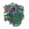
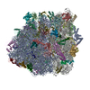
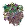
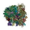
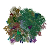
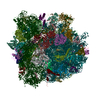
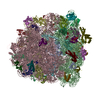
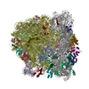
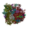
  • Deposited structure unit
  • Imaged by Jmol
  • Download
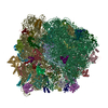
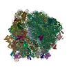
  • Superimposition on EM map
  • EMDB-8107
  • Imaged by UCSF Chimera
  • Download
Movie viewer
Structure viewerMolecule:
MolmilJmol/JSmol

Downloads & links

-
Assembly

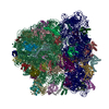
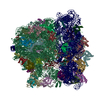
Deposited unit
B: 50S ribosomal protein L2
C: 50S ribosomal protein L3
D: 50S ribosomal protein L4
E: 50S ribosomal protein L5
F: 50S ribosomal protein L6
G: 50S ribosomal protein L9
H: 50S ribosomal protein L10
I: 50S ribosomal protein L11
J: 50S ribosomal protein L13
K: 50S ribosomal protein L14
L: 50S ribosomal protein L15
M: 50S ribosomal protein L16
N: 50S ribosomal protein L17
O: 50S ribosomal protein L18
P: 50S ribosomal protein L19
Q: 50S ribosomal protein L20
R: 50S ribosomal protein L21
S: 50S ribosomal protein L22
T: 50S ribosomal protein L23
U: 50S ribosomal protein L24
V: 50S ribosomal protein L25
W: 50S ribosomal protein L27
X: 50S ribosomal protein L28
Y: 50S ribosomal protein L29
Z: 50S ribosomal protein L30
a: 50S ribosomal protein L31
b: 50S ribosomal protein L32
c: 50S ribosomal protein L33
d: 50S ribosomal protein L34
e: 50S ribosomal protein L35
f: 50S ribosomal protein L36
g: 30S ribosomal protein S2
h: 30S ribosomal protein S3
i: 30S ribosomal protein S4
j: 30S ribosomal protein S5
k: 30S ribosomal protein S6
l: 30S ribosomal protein S7
m: 30S ribosomal protein S8
n: 30S ribosomal protein S9
o: 30S ribosomal protein S10
p: 30S ribosomal protein S11
q: 30S ribosomal protein S12
r: 30S ribosomal protein S13
s: 30S ribosomal protein S14
t: 30S ribosomal protein S15
u: 30S ribosomal protein S16
v: 30S ribosomal protein S17
w: 30S ribosomal protein S18
x: 30S ribosomal protein S19
y: 30S ribosomal protein S20
z: 30S ribosomal protein S21
1: LSU rRNA
2: SSU rRNA
3: 5S rRNA
4: E-site tRNA(Phe)
5: P-site fMet-tRNA(fMet)
6: A/T tRNA(Phe)
7: mRNA
8: GTP pyrophosphokinase
hetero molecules


Theoretical massNumber of molelcules
Total (without water)2,339,189363
Polymers2,330,96059
Non-polymers8,228304
Water362
1


  • Idetical with deposited unit
  • defined by author&software
TypeNameSymmetry operationNumber
identity operation1_5551
Buried area350280 Å2
ΔGint-5216 kcal/mol
Surface area780780 Å2
MethodPISA

-
Components

+
50S ribosomal protein ... , 31 types, 31 molecules BCDEFGHIJKLMNOPQRSTUVWXYZabcdef

#1: Protein 50S ribosomal protein L2


Mass: 29923.619 Da / Num. of mol.: 1 / Source method: isolated from a natural source / Source: (natural) Escherichia coli K-12 (bacteria) / References: UniProt: P60422
#2: Protein 50S ribosomal protein L3


Mass: 22277.535 Da / Num. of mol.: 1 / Source method: isolated from a natural source / Source: (natural) Escherichia coli K-12 (bacteria) / References: UniProt: P60438
#3: Protein 50S ribosomal protein L4


Mass: 22121.566 Da / Num. of mol.: 1 / Source method: isolated from a natural source / Source: (natural) Escherichia coli K-12 (bacteria) / References: UniProt: P60723
#4: Protein 50S ribosomal protein L5


Mass: 20333.611 Da / Num. of mol.: 1 / Source method: isolated from a natural source / Source: (natural) Escherichia coli K-12 (bacteria) / References: UniProt: P62399
#5: Protein 50S ribosomal protein L6


Mass: 18932.791 Da / Num. of mol.: 1 / Source method: isolated from a natural source / Source: (natural) Escherichia coli K-12 (bacteria) / References: UniProt: P0AG55
#6: Protein 50S ribosomal protein L9


Mass: 15789.020 Da / Num. of mol.: 1 / Source method: isolated from a natural source / Source: (natural) Escherichia coli K-12 (bacteria) / References: UniProt: P0A7R1
#7: Protein 50S ribosomal protein L10 / 50S ribosomal protein L8


Mass: 17736.596 Da / Num. of mol.: 1 / Source method: isolated from a natural source / Source: (natural) Escherichia coli K-12 (bacteria) / References: UniProt: P0A7J3
#8: Protein 50S ribosomal protein L11


Mass: 14894.362 Da / Num. of mol.: 1 / Source method: isolated from a natural source / Source: (natural) Escherichia coli K-12 (bacteria) / References: UniProt: P0A7J7
#9: Protein 50S ribosomal protein L13


Mass: 16050.606 Da / Num. of mol.: 1 / Source method: isolated from a natural source / Source: (natural) Escherichia coli K-12 (bacteria) / References: UniProt: P0AA10
#10: Protein 50S ribosomal protein L14


Mass: 13565.067 Da / Num. of mol.: 1 / Source method: isolated from a natural source / Source: (natural) Escherichia coli K-12 (bacteria) / References: UniProt: P0ADY3
#11: Protein 50S ribosomal protein L15


Mass: 15008.471 Da / Num. of mol.: 1 / Source method: isolated from a natural source / Source: (natural) Escherichia coli K-12 (bacteria) / References: UniProt: P02413
#12: Protein 50S ribosomal protein L16


Mass: 15312.269 Da / Num. of mol.: 1 / Source method: isolated from a natural source / Source: (natural) Escherichia coli K-12 (bacteria) / References: UniProt: P0ADY7
#13: Protein 50S ribosomal protein L17


Mass: 14393.657 Da / Num. of mol.: 1 / Source method: isolated from a natural source / Source: (natural) Escherichia coli K-12 (bacteria) / References: UniProt: P0AG44
#14: Protein 50S ribosomal protein L18


Mass: 12794.668 Da / Num. of mol.: 1 / Source method: isolated from a natural source / Source: (natural) Escherichia coli K-12 (bacteria) / References: UniProt: P0C018
#15: Protein 50S ribosomal protein L19


Mass: 13159.278 Da / Num. of mol.: 1 / Source method: isolated from a natural source / Source: (natural) Escherichia coli K-12 (bacteria) / References: UniProt: P0A7K6
#16: Protein 50S ribosomal protein L20


Mass: 13528.024 Da / Num. of mol.: 1 / Source method: isolated from a natural source / Source: (natural) Escherichia coli K-12 (bacteria) / References: UniProt: P0A7L3
#17: Protein 50S ribosomal protein L21


Mass: 11586.374 Da / Num. of mol.: 1 / Source method: isolated from a natural source / Source: (natural) Escherichia coli K-12 (bacteria) / References: UniProt: P0AG48
#18: Protein 50S ribosomal protein L22


Mass: 12253.359 Da / Num. of mol.: 1 / Source method: isolated from a natural source / Source: (natural) Escherichia coli K-12 (bacteria) / References: UniProt: P61175
#19: Protein 50S ribosomal protein L23


Mass: 11222.160 Da / Num. of mol.: 1 / Source method: isolated from a natural source / Source: (natural) Escherichia coli K-12 (bacteria) / References: UniProt: P0ADZ0
#20: Protein 50S ribosomal protein L24


Mass: 11339.250 Da / Num. of mol.: 1 / Source method: isolated from a natural source / Source: (natural) Escherichia coli K-12 (bacteria) / References: UniProt: P60624
#21: Protein 50S ribosomal protein L25


Mass: 10713.465 Da / Num. of mol.: 1 / Source method: isolated from a natural source / Source: (natural) Escherichia coli K-12 (bacteria) / References: UniProt: P68919
#22: Protein 50S ribosomal protein L27


Mass: 9146.540 Da / Num. of mol.: 1 / Source method: isolated from a natural source / Source: (natural) Escherichia coli K-12 (bacteria) / References: UniProt: P0A7L8
#23: Protein 50S ribosomal protein L28


Mass: 9027.551 Da / Num. of mol.: 1 / Source method: isolated from a natural source / Source: (natural) Escherichia coli K-12 (bacteria) / References: UniProt: P0A7M2
#24: Protein 50S ribosomal protein L29


Mass: 7286.464 Da / Num. of mol.: 1 / Source method: isolated from a natural source / Source: (natural) Escherichia coli K-12 (bacteria) / References: UniProt: P0A7M6
#25: Protein 50S ribosomal protein L30


Mass: 6554.820 Da / Num. of mol.: 1 / Source method: isolated from a natural source / Source: (natural) Escherichia coli K-12 (bacteria) / References: UniProt: P0AG51
#26: Protein 50S ribosomal protein L31


Mass: 7887.117 Da / Num. of mol.: 1 / Source method: isolated from a natural source / Source: (natural) Escherichia coli K-12 (bacteria) / References: UniProt: P0A7M9
#27: Protein 50S ribosomal protein L32


Mass: 6463.445 Da / Num. of mol.: 1 / Source method: isolated from a natural source / Source: (natural) Escherichia coli K-12 (bacteria) / References: UniProt: P0A7N4
#28: Protein 50S ribosomal protein L33


Mass: 6388.631 Da / Num. of mol.: 1 / Source method: isolated from a natural source / Source: (natural) Escherichia coli K-12 (bacteria) / References: UniProt: P0A7N9
#29: Protein/peptide 50S ribosomal protein L34


Mass: 5397.463 Da / Num. of mol.: 1 / Source method: isolated from a natural source / Source: (natural) Escherichia coli K-12 (bacteria) / References: UniProt: P0A7P5
#30: Protein 50S ribosomal protein L35 / Ribosomal protein A


Mass: 7313.032 Da / Num. of mol.: 1 / Source method: isolated from a natural source / Source: (natural) Escherichia coli K-12 (bacteria) / References: UniProt: P0A7Q1
#31: Protein/peptide 50S ribosomal protein L36 / Ribosomal protein B


Mass: 4377.390 Da / Num. of mol.: 1 / Source method: isolated from a natural source / Source: (natural) Escherichia coli K-12 (bacteria) / References: UniProt: P0A7Q6

-
30S ribosomal protein ... , 20 types, 20 molecules ghijklmnopqrstuvwxyz

#32: Protein 30S ribosomal protein S2


Mass: 26781.670 Da / Num. of mol.: 1 / Source method: isolated from a natural source / Source: (natural) Escherichia coli K-12 (bacteria) / References: UniProt: P0A7V0
#33: Protein 30S ribosomal protein S3


Mass: 25975.209 Da / Num. of mol.: 1 / Source method: isolated from a natural source / Source: (natural) Escherichia coli K-12 (bacteria) / References: UniProt: P0A7V3
#34: Protein 30S ribosomal protein S4


Mass: 23514.199 Da / Num. of mol.: 1 / Source method: isolated from a natural source / Source: (natural) Escherichia coli K-12 (bacteria) / References: UniProt: P0A7V8
#35: Protein 30S ribosomal protein S5


Mass: 17629.398 Da / Num. of mol.: 1 / Source method: isolated from a natural source / Source: (natural) Escherichia coli K-12 (bacteria) / References: UniProt: P0A7W1
#36: Protein 30S ribosomal protein S6


Mass: 15727.512 Da / Num. of mol.: 1 / Source method: isolated from a natural source / Source: (natural) Escherichia coli K-12 (bacteria) / References: UniProt: P02358
#37: Protein 30S ribosomal protein S7


Mass: 20055.156 Da / Num. of mol.: 1 / Source method: isolated from a natural source / Source: (natural) Escherichia coli K-12 (bacteria) / References: UniProt: P02359
#38: Protein 30S ribosomal protein S8


Mass: 14146.557 Da / Num. of mol.: 1 / Source method: isolated from a natural source / Source: (natural) Escherichia coli K-12 (bacteria) / References: UniProt: P0A7W7
#39: Protein 30S ribosomal protein S9


Mass: 14886.270 Da / Num. of mol.: 1 / Source method: isolated from a natural source / Source: (natural) Escherichia coli K-12 (bacteria) / References: UniProt: P0A7X3
#40: Protein 30S ribosomal protein S10


Mass: 11755.597 Da / Num. of mol.: 1 / Source method: isolated from a natural source / Source: (natural) Escherichia coli K-12 (bacteria) / References: UniProt: P0A7R5
#41: Protein 30S ribosomal protein S11


Mass: 13870.975 Da / Num. of mol.: 1 / Source method: isolated from a natural source / Source: (natural) Escherichia coli K-12 (bacteria) / References: UniProt: P0A7R9
#42: Protein 30S ribosomal protein S12


Mass: 13814.249 Da / Num. of mol.: 1 / Source method: isolated from a natural source / Source: (natural) Escherichia coli K-12 (bacteria) / References: UniProt: P0A7S3
#43: Protein 30S ribosomal protein S13


Mass: 13128.467 Da / Num. of mol.: 1 / Source method: isolated from a natural source / Source: (natural) Escherichia coli K-12 (bacteria) / References: UniProt: P0A7S9
#44: Protein 30S ribosomal protein S14


Mass: 11606.560 Da / Num. of mol.: 1 / Source method: isolated from a natural source / Source: (natural) Escherichia coli K-12 (bacteria) / References: UniProt: P0AG59
#45: Protein 30S ribosomal protein S15


Mass: 10290.816 Da / Num. of mol.: 1 / Source method: isolated from a natural source / Source: (natural) Escherichia coli K-12 (bacteria) / References: UniProt: P0ADZ4
#46: Protein 30S ribosomal protein S16


Mass: 9207.572 Da / Num. of mol.: 1 / Source method: isolated from a natural source / Source: (natural) Escherichia coli K-12 (bacteria) / References: UniProt: P0A7T3
#47: Protein 30S ribosomal protein S17


Mass: 9724.491 Da / Num. of mol.: 1 / Source method: isolated from a natural source / Source: (natural) Escherichia coli K-12 (bacteria) / References: UniProt: P0AG63
#48: Protein 30S ribosomal protein S18


Mass: 9063.508 Da / Num. of mol.: 1 / Source method: isolated from a natural source / Source: (natural) Escherichia coli K-12 (bacteria) / References: UniProt: P0A7T7
#49: Protein 30S ribosomal protein S19


Mass: 10455.355 Da / Num. of mol.: 1 / Source method: isolated from a natural source / Source: (natural) Escherichia coli K-12 (bacteria) / References: UniProt: P0A7U3
#50: Protein 30S ribosomal protein S20


Mass: 9708.464 Da / Num. of mol.: 1 / Source method: isolated from a natural source / Source: (natural) Escherichia coli K-12 (bacteria) / References: UniProt: P0A7U7
#51: Protein 30S ribosomal protein S21


Mass: 8524.039 Da / Num. of mol.: 1 / Source method: isolated from a natural source / Source: (natural) Escherichia coli K-12 (bacteria) / References: UniProt: P68679

-
RNA chain , 7 types, 7 molecules 1234567

#52: RNA chain LSU rRNA


Mass: 941832.625 Da / Num. of mol.: 1 / Source method: isolated from a natural source / Source: (natural) Escherichia coli K-12 (bacteria)
#53: RNA chain SSU rRNA


Mass: 497075.812 Da / Num. of mol.: 1 / Source method: isolated from a natural source / Source: (natural) Escherichia coli K-12 (bacteria)
#54: RNA chain 5S rRNA


Mass: 38177.762 Da / Num. of mol.: 1 / Source method: isolated from a natural source / Source: (natural) Escherichia coli K-12 (bacteria)
#55: RNA chain E-site tRNA(Phe)


Mass: 24485.539 Da / Num. of mol.: 1 / Source method: isolated from a natural source / Source: (natural) Escherichia coli K-12 (bacteria)
#56: RNA chain P-site fMet-tRNA(fMet)


Mass: 24893.010 Da / Num. of mol.: 1 / Source method: isolated from a natural source / Source: (natural) Escherichia coli K-12 (bacteria)
#57: RNA chain A/T tRNA(Phe)


Mass: 24707.926 Da / Num. of mol.: 1 / Source method: isolated from a natural source / Source: (natural) Escherichia coli K-12 (bacteria)
#58: RNA chain mRNA


Mass: 3146.916 Da / Num. of mol.: 1 / Source method: obtained synthetically / Source: (synth.) synthetic construct (others)

-
Protein , 1 types, 1 molecules 8

#59: Protein GTP pyrophosphokinase / (p)ppGpp synthase / ATP:GTP 3'-pyrophosphotransferase / ppGpp synthase I


Mass: 83996.617 Da / Num. of mol.: 1 / Source method: isolated from a natural source / Source: (natural) Escherichia coli K-12 (bacteria) / References: UniProt: P0AG20, GTP diphosphokinase

-
Non-polymers , 5 types, 306 molecules
#60: Chemical...
ChemComp-MG / MAGNESIUM ION


Mass: 24.305 Da / Num. of mol.: 299 / Source method: obtained synthetically / Formula: Mg
#61: Chemical ChemComp-ZN / ZINC ION


Mass: 65.409 Da / Num. of mol.: 3 / Source method: obtained synthetically / Formula: Zn
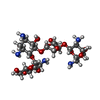
#62: Chemical ChemComp-PAR / PAROMOMYCIN / PAROMOMYCIN I / AMMINOSIDIN / CATENULIN / CRESTOMYCIN / MONOMYCIN A / NEOMYCIN E


Mass: 615.628 Da / Num. of mol.: 1 / Source method: obtained synthetically / Formula: C23H45N5O14 / Comment: Antimicrobial, medication*YM
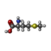
#63: Chemical ChemComp-MET / METHIONINE


Type: L-peptide linking / Mass: 149.211 Da / Num. of mol.: 1 / Source method: isolated from a natural source / Formula: C5H11NO2S
#64: Water ChemComp-HOH / water


Mass: 18.015 Da / Num. of mol.: 2 / Source method: isolated from a natural source / Formula: H2O

-
Experimental details

-
Experiment

ExperimentMethod: ELECTRON MICROSCOPY
EM experimentAggregation state: PARTICLE / 3D reconstruction method: single particle reconstruction

-
Sample preparation

ComponentName: RIBOSOME / Type: RIBOSOME / Entity ID: #1-#59 / Source: NATURAL
Source (natural)Organism: Escherichia coli K-12 (bacteria)
Buffer solutionpH: 7.5
Buffer component
IDConc.NameFormulaBuffer-ID
15 mMHepes1
250 mMKCl1
310 mMNH4Cl1
410 mMMg(OAc)21
56 mMbeta-mercaptoethanol1
SpecimenConc.: 0.1 mg/ml / Embedding applied: NO / Shadowing applied: NO / Staining applied: NO / Vitrification applied: YES / Details: This sample was monodisperse.
Specimen supportGrid material: COPPER / Grid mesh size: 400 divisions/in. / Grid type: Quantifoil R2/2
VitrificationInstrument: FEI VITROBOT MARK II / Cryogen name: ETHANE / Humidity: 100 % / Chamber temperature: 277 K / Details: Grids were blotted for 5 s

-
Electron microscopy imaging

Experimental equipment
Model: Tecnai Polara / Image courtesy: FEI Company
MicroscopyModel: FEI POLARA 300
Electron gunElectron source: FIELD EMISSION GUN / Accelerating voltage: 300 kV / Illumination mode: FLOOD BEAM
Electron lensMode: BRIGHT FIELD / Calibrated magnification: 104478 X / Nominal defocus max: 3000 nm / Nominal defocus min: 1800 nm / Alignment procedure: COMA FREE
Specimen holderCryogen: NITROGEN
Image recordingElectron dose: 35 e/Å2 / Film or detector model: OTHER
Details: FEI Falcon III Images were collected in movie-mode at 30 frames per second

-
Processing

SoftwareName: REFMAC / Version: 5.8.0124 / Classification: refinement
EM software
IDNameVersionCategoryDetails
1EMAN2.1particle selectionEMAN2 e2boxer.py was used to select particles
2EPUimage acquisitionused to collect images
4Gctf0.5CTF correction
7UCSF Chimera1.8.1model fitting
8Coot0.8.3model fitting
10RELION1.4initial Euler assignment
11RELION1.4final Euler assignment
12RELION1.4classification
13RELION1.43D reconstruction
14REFMAC5.8.0124model refinement
CTF correctionType: NONE
Particle selectionNum. of particles selected: 183615
3D reconstructionResolution: 3 Å / Resolution method: FSC 0.143 CUT-OFF / Num. of particles: 98498 / Symmetry type: POINT
Atomic model buildingB value: 112.7 / Protocol: OTHER / Space: RECIPROCAL / Target criteria: Average FSC
Atomic model buildingPDB-ID: 4YBB
Accession code: 4YBB / Source name: PDB / Type: experimental model
RefinementResolution: 3→257.28 Å / Cor.coef. Fo:Fc: 0.907 / SU B: 16.623 / SU ML: 0.301 / ESU R: 0.472
Stereochemistry target values: MAXIMUM LIKELIHOOD WITH PHASES
Details: HYDROGENS HAVE BEEN ADDED IN THE RIDING POSITIONS
RfactorNum. reflection% reflection
Rwork0.34133 --
obs0.34133 1321394 100 %
Solvent computationSolvent model: PARAMETERS FOR MASK CACLULATION
Displacement parametersBiso mean: 106.009 Å2
Baniso -1Baniso -2Baniso -3
1-0.64 Å20.13 Å2-0.01 Å2
2---0.55 Å20.26 Å2
3----0.09 Å2
Refinement stepCycle: 1 / Total: 151800
Refine LS restraints
Refine-IDTypeDev idealDev ideal targetNumber
ELECTRON MICROSCOPYr_bond_refined_d0.0050.013168776
ELECTRON MICROSCOPYr_bond_other_d0.0030.0297342
ELECTRON MICROSCOPYr_angle_refined_deg1.0611.464245806
ELECTRON MICROSCOPYr_angle_other_deg1.2962.992229327
ELECTRON MICROSCOPYr_dihedral_angle_1_deg18.2338.17626316
ELECTRON MICROSCOPYr_dihedral_angle_2_deg33.36822.262102
ELECTRON MICROSCOPYr_dihedral_angle_3_deg17.006159300
ELECTRON MICROSCOPYr_dihedral_angle_4_deg14.32115575
ELECTRON MICROSCOPYr_chiral_restr0.510.22128615
ELECTRON MICROSCOPYr_gen_planes_refined0.0090.02112835
ELECTRON MICROSCOPYr_gen_planes_other0.0030.0237007
ELECTRON MICROSCOPYr_nbd_refined
ELECTRON MICROSCOPYr_nbd_other
ELECTRON MICROSCOPYr_nbtor_refined
ELECTRON MICROSCOPYr_nbtor_other
ELECTRON MICROSCOPYr_xyhbond_nbd_refined
ELECTRON MICROSCOPYr_xyhbond_nbd_other
ELECTRON MICROSCOPYr_metal_ion_refined
ELECTRON MICROSCOPYr_metal_ion_other
ELECTRON MICROSCOPYr_symmetry_vdw_refined
ELECTRON MICROSCOPYr_symmetry_vdw_other
ELECTRON MICROSCOPYr_symmetry_hbond_refined
ELECTRON MICROSCOPYr_symmetry_hbond_other
ELECTRON MICROSCOPYr_symmetry_metal_ion_refined
ELECTRON MICROSCOPYr_symmetry_metal_ion_other
ELECTRON MICROSCOPYr_mcbond_it2.44112.94124864
ELECTRON MICROSCOPYr_mcbond_other2.44112.94124863
ELECTRON MICROSCOPYr_mcangle_it4.58319.39430982
ELECTRON MICROSCOPYr_mcangle_other4.58319.39530983
ELECTRON MICROSCOPYr_scbond_it1.79610.274143912
ELECTRON MICROSCOPYr_scbond_other1.79610.274143913
ELECTRON MICROSCOPYr_scangle_it
ELECTRON MICROSCOPYr_scangle_other3.17315.455214825
ELECTRON MICROSCOPYr_long_range_B_refined11.169611822
ELECTRON MICROSCOPYr_long_range_B_other11.169611823
ELECTRON MICROSCOPYr_rigid_bond_restr
ELECTRON MICROSCOPYr_sphericity_free
ELECTRON MICROSCOPYr_sphericity_bonded
LS refinement shellResolution: 3→3.078 Å / Total num. of bins used: 20
RfactorNum. reflection% reflection
Rwork0.968 97660 -
Rfree-0 -
obs--100 %

+
About Yorodumi

-
News

-
Feb 9, 2022. New format data for meta-information of EMDB entries

New format data for meta-information of EMDB entries

  • Version 3 of the EMDB header file is now the official format.
  • The previous official version 1.9 will be removed from the archive.

Related info.:EMDB header

External links:wwPDB to switch to version 3 of the EMDB data model

-
Aug 12, 2020. Covid-19 info

Covid-19 info

URL: https://pdbj.org/emnavi/covid19.php

New page: Covid-19 featured information page in EM Navigator.

Related info.:Covid-19 info / Mar 5, 2020. Novel coronavirus structure data

+
Mar 5, 2020. Novel coronavirus structure data

Novel coronavirus structure data

Related info.:Yorodumi Speices / Aug 12, 2020. Covid-19 info

External links:COVID-19 featured content - PDBj / Molecule of the Month (242):Coronavirus Proteases

+
Jan 31, 2019. EMDB accession codes are about to change! (news from PDBe EMDB page)

EMDB accession codes are about to change! (news from PDBe EMDB page)

  • The allocation of 4 digits for EMDB accession codes will soon come to an end. Whilst these codes will remain in use, new EMDB accession codes will include an additional digit and will expand incrementally as the available range of codes is exhausted. The current 4-digit format prefixed with “EMD-” (i.e. EMD-XXXX) will advance to a 5-digit format (i.e. EMD-XXXXX), and so on. It is currently estimated that the 4-digit codes will be depleted around Spring 2019, at which point the 5-digit format will come into force.
  • The EM Navigator/Yorodumi systems omit the EMD- prefix.

Related info.:Q: What is EMD? / ID/Accession-code notation in Yorodumi/EM Navigator

External links:EMDB Accession Codes are Changing Soon! / Contact to PDBj

+
Jul 12, 2017. Major update of PDB

Major update of PDB

  • wwPDB released updated PDB data conforming to the new PDBx/mmCIF dictionary.
  • This is a major update changing the version number from 4 to 5, and with Remediation, in which all the entries are updated.
  • In this update, many items about electron microscopy experimental information are reorganized (e.g. em_software).
  • Now, EM Navigator and Yorodumi are based on the updated data.

External links:wwPDB Remediation / Enriched Model Files Conforming to OneDep Data Standards Now Available in the PDB FTP Archive

-
Yorodumi

Thousand views of thousand structures

  • Yorodumi is a browser for structure data from EMDB, PDB, SASBDB, etc.
  • This page is also the successor to EM Navigator detail page, and also detail information page/front-end page for Omokage search.
  • The word "yorodu" (or yorozu) is an old Japanese word meaning "ten thousand". "mi" (miru) is to see.

Related info.:EMDB / PDB / SASBDB / Comparison of 3 databanks / Yorodumi Search / Aug 31, 2016. New EM Navigator & Yorodumi / Yorodumi Papers / Jmol/JSmol / Function and homology information / Changes in new EM Navigator and Yorodumi

Read more