 Movie
Movie Controller

[English] 日本語
 Yorodumi
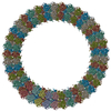
- PDB-5h3d: Helical structure of membrane tubules decorated by ACAP1 (BARPH d... -
+ Open data

- Basic information
| Entry | Database: PDB / ID: 5h3d | ||||||
|---|---|---|---|---|---|---|---|
| Title | Helical structure of membrane tubules decorated by ACAP1 (BARPH doamin) protein by cryo-electron microscopy and MD simulation | ||||||
 Components Components | Arf-GAP with coiled-coil, ANK repeat and PH domain-containing protein 1 | ||||||
 Keywords Keywords | SIGNALING PROTEIN / ACAP1 BARPH domain / membrane remodeling / molecular dynamics simulation | ||||||
| Function / homology |  Function and homology information Function and homology informationGTPase activator activity / recycling endosome membrane / protein transport / zinc ion binding / membrane Similarity search - Function | ||||||
| Biological species |  Homo sapiens (human) Homo sapiens (human) | ||||||
| Method | ELECTRON MICROSCOPY / helical reconstruction / cryo EM / Resolution: 14 Å | ||||||
 Authors Authors | Chan, C. / Pang, X.Y. / Zhang, Y. / Sun, F. / Fan, J. | ||||||
| Funding support |  China, 1items China, 1items
| ||||||
 Citation Citation | Journal: PLoS Comput Biol / Year: 2019 Title: ACAP1 assembles into an unusual protein lattice for membrane deformation through multiple stages. Authors: Chun Chan / Xiaoyun Pang / Yan Zhang / Tongxin Niu / Shengjiang Yang / Daohui Zhao / Jian Li / Lanyuan Lu / Victor W Hsu / Jian Zhou / Fei Sun / Jun Fan /   Abstract: Studies on the Bin-Amphiphysin-Rvs (BAR) domain have advanced a fundamental understanding of how proteins deform membrane. We previously showed that a BAR domain in tandem with a Pleckstrin Homology ...Studies on the Bin-Amphiphysin-Rvs (BAR) domain have advanced a fundamental understanding of how proteins deform membrane. We previously showed that a BAR domain in tandem with a Pleckstrin Homology (PH domain) underlies the assembly of ACAP1 (Arfgap with Coil-coil, Ankryin repeat, and PH domain I) into an unusual lattice structure that also uncovers a new paradigm for how a BAR protein deforms membrane. Here, we initially pursued computation-based refinement of the ACAP1 lattice to identify its critical protein contacts. Simulation studies then revealed how ACAP1, which dimerizes into a symmetrical structure in solution, is recruited asymmetrically to the membrane through dynamic behavior. We also pursued electron microscopy (EM)-based structural studies, which shed further insight into the dynamic nature of the ACAP1 lattice assembly. As ACAP1 is an unconventional BAR protein, our findings broaden the understanding of the mechanistic spectrum by which proteins assemble into higher-ordered structures to achieve membrane deformation. | ||||||
| History |
|
- Structure visualization
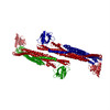
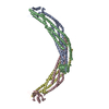
| Movie |
Movie viewer |
|---|---|
| Structure viewer | Molecule: MolmilJmol/JSmol |
- Downloads & links
-Download
| PDBx/mmCIF format | 5h3d.cif.gz | 480 KB | Display | PDBx/mmCIF format |
|---|---|---|---|---|
| PDB format | pdb5h3d.ent.gz | 390.6 KB | Display | PDB format |
| PDBx/mmJSON format | 5h3d.json.gz | Tree view | PDBx/mmJSON format | |
| Others |  Other downloads Other downloads |
-Validation report
| Arichive directory | https://data.pdbj.org/pub/pdb/validation_reports/h3/5h3dftp://data.pdbj.org/pub/pdb/validation_reports/h3/5h3d | HTTPS FTP |
|---|
-Related structure data
| Related structure data | 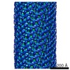 2546M M: map data used to model this data |
|---|---|
| Similar structure data |
-Links
 PDBj
PDBj

- Assembly
| Deposited unit | 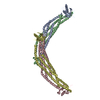
|
|---|---|
| 1 | x 9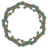
|
| 2 |
|
| 3 | 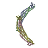
|
| Symmetry | Point symmetry: (Schoenflies symbol: C3 (3 fold cyclic)) Helical symmetry: (Circular symmetry: 3 / Dyad axis: no / N subunits divisor: 1 / Num. of operations: 9 / Rise per n subunits: 23.198 Å / Rotation per n subunits: 42.693 °) |
-Components
| #1: Protein | Mass: 43334.348 Da / Num. of mol.: 4 Source method: isolated from a genetically manipulated source Source: (gene. exp.) Homo sapiens (human) / Gene: ACAP1, CENTB1, KIAA0050 / Production host:  |
|---|
-Experimental details
-Experiment
| Experiment | Method: ELECTRON MICROSCOPY |
|---|---|
| EM experiment | Aggregation state: HELICAL ARRAY / 3D reconstruction method: helical reconstruction |
- Sample preparation
| Component | Name: Helical structure of ACAP1 BARPH domain on membrane tubules Type: COMPLEX / Entity ID: all / Source: RECOMBINANT | ||||||||||||
|---|---|---|---|---|---|---|---|---|---|---|---|---|---|
| Molecular weight | Experimental value: NO | ||||||||||||
| Source (natural) | Organism: Homo sapiens (human) | ||||||||||||
| Source (recombinant) | Organism: | ||||||||||||
| Buffer solution | pH: 7.4 / Details: 50mM HEPES, pH7.4, 100mM NaCl, pH 7.4 | ||||||||||||
| Buffer component |
| ||||||||||||
| Specimen | Conc.: 4 mg/ml / Embedding applied: NO / Shadowing applied: NO / Staining applied: NO / Vitrification applied: YES Details: The ACAP1 BAR-PH protein (4 mg/ml) was incubated with liposomes(2 mg/ml) | ||||||||||||
| Specimen support | Grid material: COPPER / Grid mesh size: 300 divisions/in. / Grid type: Homemade | ||||||||||||
| Vitrification | Instrument: FEI VITROBOT MARK IV / Cryogen name: ETHANE / Humidity: 100 % / Chamber temperature: 289 K |
- Electron microscopy imaging
| Experimental equipment |  Model: Titan Krios / Image courtesy: FEI Company |
|---|---|
| Microscopy | Model: FEI TITAN KRIOS |
| Electron gun | Electron source:  FIELD EMISSION GUN / Accelerating voltage: 300 kV / Illumination mode: OTHER FIELD EMISSION GUN / Accelerating voltage: 300 kV / Illumination mode: OTHER |
| Electron lens | Mode: BRIGHT FIELD / Nominal magnification: 75000 X / Nominal defocus max: 3500 nm / Nominal defocus min: 2500 nm / Cs: 2.7 mm |
| Specimen holder | Cryogen: NITROGEN / Specimen holder model: FEI TITAN KRIOS AUTOGRID HOLDER |
| Image recording | Electron dose: 20 e/Å2 / Film or detector model: GATAN ULTRASCAN 4000 (4k x 4k) |
- Processing
| EM software | Name: SerialEM / Category: image acquisition |
|---|---|
| CTF correction | Type: PHASE FLIPPING ONLY |
| Helical symmerty | Angular rotation/subunit: 42.72 ° / Axial rise/subunit: 23.2 Å / Axial symmetry: C3 |
| 3D reconstruction | Method: HELICAL / Resolution: 14 Å / Resolution method: FSC 0.143 CUT-OFF / Num. of particles: 304 / Symmetry type: HELICAL |
| Atomic model building | Protocol: FLEXIBLE FIT / Space: REAL / Target criteria: cross correlation |
