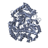
Movie
Movie Controller

[English] 日本語
 Yorodumi
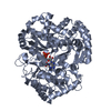
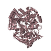
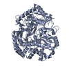
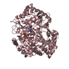
- PDB-5fj6: Structure of the P2 polymerase inside in vitro assembled bacterio... -
+ Open data

- Basic information
| Entry | Database: PDB / ID: 5fj6 | ||||||
|---|---|---|---|---|---|---|---|
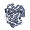
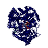
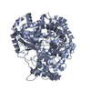
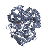
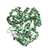
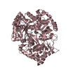
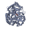
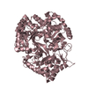
| Title | Structure of the P2 polymerase inside in vitro assembled bacteriophage phi6 polymerase complex | ||||||
 Components Components | RNA-DIRECTED RNA POLYMERASE | ||||||
 Keywords Keywords | TRANSCRIPTION / BACTERIOPHAGE PHI6 / POLYMERASE COMPLEX / P2 / POLYMERASE | ||||||
| Function / homology |  Function and homology information Function and homology informationRNA uridylyltransferase activity / virion component / RNA-directed RNA polymerase / viral RNA genome replication / nucleotide binding / RNA-directed RNA polymerase activity / DNA-templated transcription / RNA binding / metal ion binding Similarity search - Function | ||||||
| Biological species |  PSEUDOMONAS PHAGE PHI6 (virus) PSEUDOMONAS PHAGE PHI6 (virus) | ||||||
| Method | ELECTRON MICROSCOPY / single particle reconstruction / cryo EM / Resolution: 7.9 Å | ||||||
 Authors Authors | Ilca, S. / Kotecha, A. / Sun, X. / Poranen, M.P. / Stuart, D.I. / Huiskonen, J.T. | ||||||
 Citation Citation | Journal: Nat Commun / Year: 2015 Title: Localized reconstruction of subunits from electron cryomicroscopy images of macromolecular complexes. Authors: Serban L Ilca / Abhay Kotecha / Xiaoyu Sun / Minna M Poranen / David I Stuart / Juha T Huiskonen /   Abstract: Electron cryomicroscopy can yield near-atomic resolution structures of highly ordered macromolecular complexes. Often however some subunits bind in a flexible manner, have different symmetry from the ...Electron cryomicroscopy can yield near-atomic resolution structures of highly ordered macromolecular complexes. Often however some subunits bind in a flexible manner, have different symmetry from the rest of the complex, or are present in sub-stoichiometric amounts, limiting the attainable resolution. Here we report a general method for the localized three-dimensional reconstruction of such subunits. After determining the particle orientations, local areas corresponding to the subunits can be extracted and treated as single particles. We demonstrate the method using three examples including a flexible assembly and complexes harbouring subunits with either partial occupancy or mismatched symmetry. Most notably, the method allows accurate fitting of the monomeric RNA-dependent RNA polymerase bound at the threefold axis of symmetry inside a viral capsid, revealing for the first time its exact orientation and interactions with the capsid proteins. Localized reconstruction is expected to provide novel biological insights in a range of challenging biological systems. | ||||||
| History |
|
- Structure visualization
| Movie |
Movie viewer |
|---|---|
| Structure viewer | Molecule: MolmilJmol/JSmol |
- Downloads & links
-Download
| PDBx/mmCIF format | 5fj6.cif.gz | 143.4 KB | Display | PDBx/mmCIF format |
|---|---|---|---|---|
| PDB format | pdb5fj6.ent.gz | 112.4 KB | Display | PDB format |
| PDBx/mmJSON format | 5fj6.json.gz | Tree view | PDBx/mmJSON format | |
| Others |  Other downloads Other downloads |
-Validation report
| Summary document | 5fj6_validation.pdf.gz | 862.7 KB | Display | wwPDB validaton report |
|---|---|---|---|---|
| Full document | 5fj6_full_validation.pdf.gz | 911.1 KB | Display | |
| Data in XML | 5fj6_validation.xml.gz | 32.7 KB | Display | |
| Data in CIF | 5fj6_validation.cif.gz | 47.6 KB | Display | |
| Arichive directory | https://data.pdbj.org/pub/pdb/validation_reports/fj/5fj6ftp://data.pdbj.org/pub/pdb/validation_reports/fj/5fj6 | HTTPS FTP |
-Related structure data
| Related structure data | 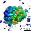 3186MC 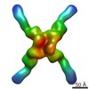 3183C 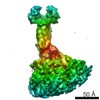 3184C 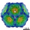 3185C 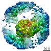 3187C 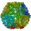 5fj5C 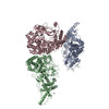 5fj7C C: citing same article ( M: map data used to model this data |
|---|---|
| Similar structure data |


-Links
 PDBj
PDBj- Assembly
| Deposited unit | 
|
|---|---|
| 1 |
|
-Components
| #1: Protein | Mass: 74903.203 Da / Num. of mol.: 1 Source method: isolated from a genetically manipulated source Source: (gene. exp.) PSEUDOMONAS PHAGE PHI6 (virus) / Production host:  PSEUDOMONAS SYRINGAE (bacteria) / References: UniProt: P11124 PSEUDOMONAS SYRINGAE (bacteria) / References: UniProt: P11124 |
|---|---|
| #2: Chemical | ChemComp-MN /   Mass: 54.938 Da / Num. of mol.: 1 / Source method: obtained synthetically / Formula: Mn Mass: 54.938 Da / Num. of mol.: 1 / Source method: obtained synthetically / Formula: Mn |
-Experimental details
-Experiment
| Experiment | Method: ELECTRON MICROSCOPY |
|---|---|
| EM experiment | Aggregation state: PARTICLE / 3D reconstruction method: single particle reconstruction |
- Sample preparation
| Component | Name: BACTERIOPHAGE PHI6 POLYMERASE COMPLEX ASSEMBLED IN VITRO FROM PURIFIED PROTEINS P1, P2, AND P4 Type: COMPLEX |
|---|---|
| Buffer solution | Name: 20 MM TRIS / pH: 8 / Details: 20 MM TRIS |
| Specimen | Conc.: 2.4 mg/ml / Embedding applied: NO / Shadowing applied: NO / Staining applied: NO / Vitrification applied: YES |
| Specimen support | Details: HOLEY CARBON |
| Vitrification | Instrument: FEI VITROBOT MARK IV / Cryogen name: ETHANE Details: VITRIFICATION 1 -- CRYOGEN- ETHANE, TEMPERATURE- 120, INSTRUMENT- FEI VITROBOT MARK IV, METHOD- BLOT 4 SECONDS BEFORE PLUNGING, |
- Electron microscopy imaging
| Experimental equipment |  Model: Tecnai F30 / Image courtesy: FEI Company |
|---|---|
| Microscopy | Model: FEI TECNAI F30 / Date: Jun 12, 2014 / Details: DOSE RATE 6-8 E- PER PIX PER S |
| Electron gun | Electron source:  FIELD EMISSION GUN / Accelerating voltage: 300 kV / Illumination mode: FLOOD BEAM FIELD EMISSION GUN / Accelerating voltage: 300 kV / Illumination mode: FLOOD BEAM |
| Electron lens | Mode: BRIGHT FIELD / Nominal magnification: 160000 X / Calibrated magnification: 37037 X / Nominal defocus max: 2600 nm / Nominal defocus min: 1100 nm / Cs: 2 mm |
| Specimen holder | Temperature: 81 K |
| Image recording | Electron dose: 16 e/Å2 / Film or detector model: GATAN K2 (4k x 4k) |
| Image scans | Num. digital images: 834 |
- Processing
| EM software |
| ||||||||||||
|---|---|---|---|---|---|---|---|---|---|---|---|---|---|
| CTF correction | Details: EACH PARTICLE | ||||||||||||
| Symmetry | Point symmetry: C1 (asymmetric) | ||||||||||||
| 3D reconstruction | Method: LOCALIZED RECONSTRUCTION / Resolution: 7.9 Å / Num. of particles: 43216 / Nominal pixel size: 1.3 Å / Actual pixel size: 1.35 Å / Magnification calibration: ATOMIC MODEL Details: SUBMISSION BASED ON EXPERIMENTAL DATA FROM EMDB EMD-3186. (DEPOSITION ID: 13857). Symmetry type: POINT | ||||||||||||
| Atomic model building | Protocol: RIGID BODY FIT / Space: REAL / Details: METHOD--RIGID BODY REFINEMENT PROTOCOL--X-RAY | ||||||||||||
| Atomic model building | PDB-ID: 1HHS Accession code: 1HHS / Source name: PDB / Type: experimental model | ||||||||||||
| Refinement | Highest resolution: 7.9 Å | ||||||||||||
| Refinement step | Cycle: LAST / Highest resolution: 7.9 Å
|