 Movie
Movie Controller

[English] 日本語
 Yorodumi
- PDB-5a7u: Single-particle cryo-EM of co-translational folded adr1 domain in... -
+ Open data

- Basic information
| Entry | Database: PDB / ID: 5a7u | ||||||
|---|---|---|---|---|---|---|---|
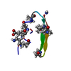
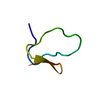
| Title | Single-particle cryo-EM of co-translational folded adr1 domain inside the E. coli ribosome exit tunnel. | ||||||
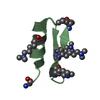
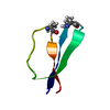
 Components Components | REGULATORY PROTEIN ADR1 | ||||||
 Keywords Keywords | TRANSLATION / PROTEIN FOLDING / RIBOSOME / ZINC FINGER / SECM / TRANSLATIONAL ARREST PEPTIDE / CRYO-EM / SINGLE- MOLECULE STUDIES | ||||||
| Function / homology |  Function and homology information Function and homology informationpositive regulation of peroxisome organization / peroxisome organization / positive regulation of fatty acid beta-oxidation / TFIIB-class transcription factor binding / TFIID-class transcription factor complex binding / cellular response to ethanol / chromatin organization / sequence-specific DNA binding / molecular adaptor activity / nucleic acid binding ...positive regulation of peroxisome organization / peroxisome organization / positive regulation of fatty acid beta-oxidation / TFIIB-class transcription factor binding / TFIID-class transcription factor complex binding / cellular response to ethanol / chromatin organization / sequence-specific DNA binding / molecular adaptor activity / nucleic acid binding / RNA polymerase II-specific DNA-binding transcription factor binding / DNA-binding transcription factor activity, RNA polymerase II-specific / transcription coactivator activity / RNA polymerase II cis-regulatory region sequence-specific DNA binding / regulation of transcription by RNA polymerase II / chromatin / positive regulation of transcription by RNA polymerase II / zinc ion binding / nucleus / cytoplasm / cytosol Similarity search - Function | ||||||
| Biological species |  | ||||||
| Method | ELECTRON MICROSCOPY / single particle reconstruction / cryo EM / Resolution: 4.8 Å | ||||||
 Authors Authors | Nilsson, O.B. / Hedman, R. / Marino, J. / Wickles, S. / Bischoff, L. / Johansson, M. / Muller-Lucks, A. / Trovato, F. / Puglisi, J.D. / O'Brien, E. ...Nilsson, O.B. / Hedman, R. / Marino, J. / Wickles, S. / Bischoff, L. / Johansson, M. / Muller-Lucks, A. / Trovato, F. / Puglisi, J.D. / O'Brien, E. / Beckmann, R. / von Heijne, G. | ||||||
 Citation Citation | Journal: Cell Rep / Year: 2015 Title: Cotranslational Protein Folding inside the Ribosome Exit Tunnel. Authors: Ola B Nilsson / Rickard Hedman / Jacopo Marino / Stephan Wickles / Lukas Bischoff / Magnus Johansson / Annika Müller-Lucks / Fabio Trovato / Joseph D Puglisi / Edward P O'Brien / Roland ...Authors: Ola B Nilsson / Rickard Hedman / Jacopo Marino / Stephan Wickles / Lukas Bischoff / Magnus Johansson / Annika Müller-Lucks / Fabio Trovato / Joseph D Puglisi / Edward P O'Brien / Roland Beckmann / Gunnar von Heijne /   Abstract: At what point during translation do proteins fold? It is well established that proteins can fold cotranslationally outside the ribosome exit tunnel, whereas studies of folding inside the exit tunnel ...At what point during translation do proteins fold? It is well established that proteins can fold cotranslationally outside the ribosome exit tunnel, whereas studies of folding inside the exit tunnel have so far detected only the formation of helical secondary structure and collapsed or partially structured folding intermediates. Here, using a combination of cotranslational nascent chain force measurements, inter-subunit fluorescence resonance energy transfer studies on single translating ribosomes, molecular dynamics simulations, and cryoelectron microscopy, we show that a small zinc-finger domain protein can fold deep inside the vestibule of the ribosome exit tunnel. Thus, for small protein domains, the ribosome itself can provide the kind of sheltered folding environment that chaperones provide for larger proteins. | ||||||
| History |
|
- Structure visualization
| Movie |
Movie viewer |
|---|---|
| Structure viewer | Molecule: MolmilJmol/JSmol |
- Downloads & links
-Download
| PDBx/mmCIF format | 5a7u.cif.gz | 21.4 KB | Display | PDBx/mmCIF format |
|---|---|---|---|---|
| PDB format | pdb5a7u.ent.gz | 12 KB | Display | PDB format |
| PDBx/mmJSON format | 5a7u.json.gz | Tree view | PDBx/mmJSON format | |
| Others |  Other downloads Other downloads |
-Validation report
| Arichive directory | https://data.pdbj.org/pub/pdb/validation_reports/a7/5a7uftp://data.pdbj.org/pub/pdb/validation_reports/a7/5a7u | HTTPS FTP |
|---|
-Related structure data
| Related structure data | 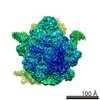 3079MC M: map data used to model this data C: citing same article ( |
|---|---|
| Similar structure data |


-Links
 PDBj
PDBj


- Assembly
| Deposited unit | 
|
|---|---|
| 1 |
|
-Components
| #1: Protein/peptide | Mass: 3406.024 Da / Num. of mol.: 1 / Fragment: RESIDUES 130-158 Source method: isolated from a genetically manipulated source Source: (gene. exp.) Production host:  |
|---|---|
| #2: Chemical | ChemComp-ZN /   Mass: 65.409 Da / Num. of mol.: 1 / Source method: obtained synthetically / Formula: Zn Mass: 65.409 Da / Num. of mol.: 1 / Source method: obtained synthetically / Formula: Zn |
-Experimental details
-Experiment
| Experiment | Method: ELECTRON MICROSCOPY |
|---|---|
| EM experiment | Aggregation state: PARTICLE / 3D reconstruction method: single particle reconstruction |
- Sample preparation
| Component | Name: ADR1 DOMAIN COTRANSLATIONALLY FOLDED INSIDE THE E. COLI RIBOSOME EXIT TUNNEL Type: RIBOSOME |
|---|---|
| Buffer solution | Name: 20 MM HEPES PH 7.2 , 50 MM KOAC, 5 MM MG OAC2, 0.03% DDM, 50 MICROM ZNCL2, 125 MM SUCROSE. pH: 7.2 Details: 20 MM HEPES PH 7.2 , 50 MM KOAC, 5 MM MG OAC2, 0.03% DDM, 50 MICROM ZNCL2, 125 MM SUCROSE. |
| Specimen | Embedding applied: NO / Shadowing applied: NO / Staining applied: NO / Vitrification applied: YES |
| Specimen support | Details: CARBON |
| Vitrification | Instrument: FEI VITROBOT MARK IV / Cryogen name: ETHANE Details: VITRIFICATION 1 -- CRYOGEN- ETHANE, INSTRUMENT- FEI VITROBOT MARK IV, |
- Electron microscopy imaging
| Experimental equipment |  Model: Titan Krios / Image courtesy: FEI Company |
|---|---|
| Microscopy | Model: FEI TITAN KRIOS / Date: Apr 13, 2015 |
| Electron gun | Electron source:  FIELD EMISSION GUN / Accelerating voltage: 300 kV / Illumination mode: SPOT SCAN FIELD EMISSION GUN / Accelerating voltage: 300 kV / Illumination mode: SPOT SCAN |
| Electron lens | Mode: BRIGHT FIELD / Nominal defocus max: 3200 nm / Nominal defocus min: 1000 nm |
| Image recording | Electron dose: 5 e/Å2 / Film or detector model: FEI FALCON II (4k x 4k) |
| Image scans | Num. digital images: 2000 |
| Radiation wavelength | Relative weight: 1 |
- Processing
| EM software |
| ||||||||||||
|---|---|---|---|---|---|---|---|---|---|---|---|---|---|
| CTF correction | Details: MICROGRAPH | ||||||||||||
| Symmetry | Point symmetry: C1 (asymmetric) | ||||||||||||
| 3D reconstruction | Resolution: 4.8 Å / Num. of particles: 151900 / Nominal pixel size: 3.7 Å / Actual pixel size: 3.7 Å Details: SUBMISSION BASED ON EXPERIMENTAL DATA FROM EMDB EMD-3079. (DEPOSITION ID: 13581). Symmetry type: POINT | ||||||||||||
| Atomic model building | Protocol: RIGID BODY FIT / Space: REAL / Details: METHOD--RIGID BODY REFINEMENT PROTOCOL--NMR | ||||||||||||
| Atomic model building | PDB-ID: 2ADR Accession code: 2ADR / Source name: PDB / Type: experimental model | ||||||||||||
| Refinement | Highest resolution: 4.8 Å | ||||||||||||
| Refinement step | Cycle: LAST / Highest resolution: 4.8 Å
|

