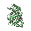
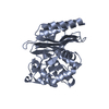
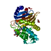
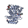
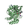
[English] 日本語
Yorodumi
- PDB-5top: Atomic Resolution X-Ray Crystal Structure of a Ruthenocene Conjug... -

+
Open data


ID or keywords:

Loading...

-
Basic information

Entry
Database: PDB / ID: 5top
TitleAtomic Resolution X-Ray Crystal Structure of a Ruthenocene Conjugated Beta-Lactam Antibiotic in Complex with CTX-M-14 S70G Beta-Lactamase
ComponentsBeta-lactamase
KeywordsHYDROLASE/ANTIBIOTIC / Beta-Lactamase / Organometallic / Ruthenocene / CTX-M-14 / HYDROLASE-ANTIBIOTIC complex
Function / homology
Function and homology information


beta-lactam antibiotic catabolic process / beta-lactamase activity / beta-lactamase / response to antibiotic
Similarity search - Function
Beta-lactamase, class-A active site / Beta-lactamase class-A active site. / Beta-lactamase class A, catalytic domain / Beta-lactamase enzyme family / Beta-lactamase, class-A / Beta-lactamase / DD-peptidase/beta-lactamase superfamily / Beta-lactamase/transpeptidase-like / 3-Layer(aba) Sandwich / Alpha Beta
Similarity search - Domain/homology
ruthenocene / Chem-JSC / Chem-JSE / : / RUTHENIUM ION / Beta-lactamase
Similarity search - Component
Biological speciesEscherichia coli (E. coli)
MethodX-RAY DIFFRACTION / SYNCHROTRON / MOLECULAR REPLACEMENT / Resolution: 1.18 Å
AuthorsLewandowski, E.M. / Chen, Y.
Funding support United States, Poland, 2items
OrganizationGrant numberCountry
National Institutes of Health/National Institute Of Allergy and Infectious Diseases (NIH/NIAID)AI103158 United States
Polish National Science CentreDEC-2013/11/B/ST5/00997 Poland
CitationJournal: FEBS J. / Year: 2018
Title: Mechanisms of proton relay and product release by Class A beta-lactamase at ultrahigh resolution.
Authors: Lewandowski, E.M. / Lethbridge, K.G. / Sanishvili, R. / Skiba, J. / Kowalski, K. / Chen, Y.
History
DepositionOct 18, 2016Deposition site: RCSB / Processing site: RCSB
Revision 1.0Nov 8, 2017Provider: repository / Type: Initial release
Revision 1.1Nov 15, 2017Group: Database references / Category: citation
Item: _citation.journal_abbrev / _citation.journal_id_ISSN ..._citation.journal_abbrev / _citation.journal_id_ISSN / _citation.pdbx_database_id_PubMed / _citation.title
Revision 1.2Jan 17, 2018Group: Database references / Category: citation
Item: _citation.journal_volume / _citation.page_first ..._citation.journal_volume / _citation.page_first / _citation.page_last / _citation.title / _citation.year
Revision 2.0Dec 11, 2019Group: Author supporting evidence / Polymer sequence / Category: entity_poly / pdbx_audit_support
Item: _entity_poly.pdbx_seq_one_letter_code_can / _pdbx_audit_support.funding_organization
Revision 2.1Oct 4, 2023Group: Data collection / Database references ...Data collection / Database references / Derived calculations / Refinement description
Category: chem_comp_atom / chem_comp_bond ...chem_comp_atom / chem_comp_bond / database_2 / pdbx_initial_refinement_model / pdbx_struct_conn_angle / struct_conn
Item: _database_2.pdbx_DOI / _database_2.pdbx_database_accession ..._database_2.pdbx_DOI / _database_2.pdbx_database_accession / _pdbx_struct_conn_angle.ptnr1_auth_asym_id / _pdbx_struct_conn_angle.ptnr1_auth_comp_id / _pdbx_struct_conn_angle.ptnr1_auth_seq_id / _pdbx_struct_conn_angle.ptnr1_label_alt_id / _pdbx_struct_conn_angle.ptnr1_label_asym_id / _pdbx_struct_conn_angle.ptnr1_label_comp_id / _pdbx_struct_conn_angle.ptnr1_label_seq_id / _pdbx_struct_conn_angle.ptnr2_auth_asym_id / _pdbx_struct_conn_angle.ptnr2_auth_comp_id / _pdbx_struct_conn_angle.ptnr2_auth_seq_id / _pdbx_struct_conn_angle.ptnr2_label_asym_id / _pdbx_struct_conn_angle.ptnr2_label_atom_id / _pdbx_struct_conn_angle.ptnr2_label_comp_id / _pdbx_struct_conn_angle.ptnr3_auth_seq_id / _pdbx_struct_conn_angle.ptnr3_label_alt_id / _pdbx_struct_conn_angle.ptnr3_symmetry / _pdbx_struct_conn_angle.value / _struct_conn.pdbx_dist_value / _struct_conn.pdbx_ptnr2_label_alt_id / _struct_conn.ptnr1_auth_asym_id / _struct_conn.ptnr1_auth_comp_id / _struct_conn.ptnr1_auth_seq_id / _struct_conn.ptnr1_label_asym_id / _struct_conn.ptnr1_label_atom_id / _struct_conn.ptnr1_label_comp_id / _struct_conn.ptnr1_label_seq_id / _struct_conn.ptnr2_auth_asym_id / _struct_conn.ptnr2_auth_comp_id / _struct_conn.ptnr2_auth_seq_id / _struct_conn.ptnr2_label_asym_id / _struct_conn.ptnr2_label_atom_id / _struct_conn.ptnr2_label_comp_id / _struct_conn.ptnr2_symmetry

-
Structure visualization

Structure viewerMolecule:
MolmilJmol/JSmol

Downloads & links

-
Assembly

Deposited unit
A: Beta-lactamase
B: Beta-lactamase
hetero molecules


Theoretical massNumber of molelcules
Total (without water)58,13511
Polymers55,9072
Non-polymers2,2289
Water10,413578
1
A: Beta-lactamase
hetero molecules


Theoretical massNumber of molelcules
Total (without water)29,1715
Polymers27,9531
Non-polymers1,2174
Water181
TypeNameSymmetry operationNumber
identity operation1_555x,y,z1
2
B: Beta-lactamase
hetero molecules


Theoretical massNumber of molelcules
Total (without water)28,9646
Polymers27,9531
Non-polymers1,0115
Water181
TypeNameSymmetry operationNumber
identity operation1_555x,y,z1
Unit cell
Length a, b, c (Å)45.093, 106.821, 47.933
Angle α, β, γ (deg.)90.00, 101.66, 90.00
Int Tables number4
Space group name H-MP1211

-
Components

-
Protein , 1 types, 2 molecules AB

#1: Protein Beta-lactamase / CTX-M-14 Class A Beta-Lactamase


Mass: 27953.488 Da / Num. of mol.: 2 / Mutation: S70G
Source method: isolated from a genetically manipulated source
Source: (gene. exp.) Escherichia coli (E. coli) / Gene: CTX-M-14 / Production host: Escherichia coli (E. coli) / References: UniProt: H6UQI0, beta-lactamase

-
Non-polymers , 6 types, 587 molecules

#2: Chemical ChemComp-K / POTASSIUM ION


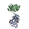
Mass: 39.098 Da / Num. of mol.: 2 / Source method: obtained synthetically / Formula: K
#3: Chemical ChemComp-JSE / [(1,2,3,4,5-eta)-1-(4-{[carboxy(4-carboxy-5,5-dimethyl-1,3-thiazolidin-2-yl)methyl]amino}-4-oxobutanoyl)cyclopentadienyl][(1,2,3,4,5-eta)-cyclopentadienyl]ruthenium


Mass: 538.515 Da / Num. of mol.: 2 / Source method: obtained synthetically / Formula: C22H17N2O6RuS
#4: Chemical ChemComp-RU / RUTHENIUM ION


Mass: 101.070 Da / Num. of mol.: 3 / Source method: obtained synthetically / Formula: Ru
#5: Chemical ChemComp-JSC / [(1,2,3,4,5-eta)-1-(4-{[carboxy(4-carboxy-5,5-dimethyl-1,3-thiazolidin-2-yl)methyl]amino}-4-oxobutanoyl)cyclopentadienyl][(1,2,3,4,5-eta)-cyclopentadienyl]ruthenium


Mass: 538.515 Da / Num. of mol.: 1 / Source method: obtained synthetically / Formula: C22H17N2O6RuS
#6: Chemical ChemComp-7G4 / ruthenocene / bis[(1,2,3,4,5-eta)-cyclopentadienyl]ruthenium


Mass: 231.256 Da / Num. of mol.: 1 / Source method: obtained synthetically / Formula: C10H10Ru
#7: Water ChemComp-HOH / water


Mass: 18.015 Da / Num. of mol.: 578 / Source method: isolated from a natural source / Formula: H2O

-
Experimental details

-
Experiment

ExperimentMethod: X-RAY DIFFRACTION / Number of used crystals: 1

-
Sample preparation

CrystalDensity Matthews: 2.01 Å3/Da / Density % sol: 38.9 %
Crystal growTemperature: 293.15 K / Method: vapor diffusion, hanging drop / pH: 7.9 / Details: potassium phosphate

-
Data collection

DiffractionMean temperature: 100 K
Diffraction sourceSource: SYNCHROTRON / Site: APS / Beamline: 22-ID / Wavelength: 1 Å
DetectorType: RAYONIX MX300-HS / Detector: CCD / Date: Apr 1, 2015
RadiationMonochromator: double crystal Si(111) / Protocol: SINGLE WAVELENGTH / Monochromatic (M) / Laue (L): M / Scattering type: x-ray
Radiation wavelengthWavelength: 1 Å / Relative weight: 1
ReflectionResolution: 1.18→50 Å / Num. obs: 133593 / % possible obs: 97.7 % / Redundancy: 3.7 % / Rmerge(I) obs: 0.036 / Net I/σ(I): 25.8
Reflection shellResolution: 1.18→1.2 Å / Redundancy: 3.1 % / Mean I/σ(I) obs: 4.3 / % possible all: 88.6

-
Processing

Software
NameVersionClassification
REFMAC5.8.0151refinement
HKL-2000data reduction
SCALEPACKdata scaling
PHASERphasing
RefinementMethod to determine structure: MOLECULAR REPLACEMENT
Starting model: PDB entry 4XXR
Resolution: 1.18→50 Å / Cor.coef. Fo:Fc: 0.981 / Cor.coef. Fo:Fc free: 0.976 / SU B: 0.942 / SU ML: 0.02 / Cross valid method: THROUGHOUT / ESU R: 0.038 / ESU R Free: 0.035 / Details: HYDROGENS HAVE BEEN ADDED IN THE RIDING POSITIONS
RfactorNum. reflection% reflectionSelection details
Rfree0.14633 7118 5.1 %RANDOM
Rwork0.12756 ---
obs0.12848 133593 96.77 %-
Solvent computationIon probe radii: 0.8 Å / Shrinkage radii: 0.8 Å / VDW probe radii: 1.2 Å
Displacement parametersBiso mean: 16.345 Å2
Baniso -1Baniso -2Baniso -3
1-0.44 Å20 Å2-0.55 Å2
2---0.47 Å20 Å2
3---0.24 Å2
Refinement stepCycle: LAST / Resolution: 1.18→50 Å
ProteinNucleic acidLigandSolventTotal
Num. atoms3903 0 112 578 4593
Refine LS restraints
Refine-IDTypeDev idealDev ideal targetNumber
X-RAY DIFFRACTIONr_bond_refined_d0.0070.0194731
X-RAY DIFFRACTIONr_bond_other_d0.0010.024477
X-RAY DIFFRACTIONr_angle_refined_deg1.3452.0016457
X-RAY DIFFRACTIONr_angle_other_deg0.687310292
X-RAY DIFFRACTIONr_dihedral_angle_1_deg6.9835630
X-RAY DIFFRACTIONr_dihedral_angle_2_deg40.42624.433194
X-RAY DIFFRACTIONr_dihedral_angle_3_deg14.34215775
X-RAY DIFFRACTIONr_dihedral_angle_4_deg14.8041539
X-RAY DIFFRACTIONr_chiral_restr0.0850.2730
X-RAY DIFFRACTIONr_gen_planes_refined0.0110.0215662
X-RAY DIFFRACTIONr_gen_planes_other0.0040.021081
X-RAY DIFFRACTIONr_nbd_refined
X-RAY DIFFRACTIONr_nbd_other
X-RAY DIFFRACTIONr_nbtor_refined
X-RAY DIFFRACTIONr_nbtor_other
X-RAY DIFFRACTIONr_xyhbond_nbd_refined
X-RAY DIFFRACTIONr_xyhbond_nbd_other
X-RAY DIFFRACTIONr_metal_ion_refined
X-RAY DIFFRACTIONr_metal_ion_other
X-RAY DIFFRACTIONr_symmetry_vdw_refined
X-RAY DIFFRACTIONr_symmetry_vdw_other
X-RAY DIFFRACTIONr_symmetry_hbond_refined
X-RAY DIFFRACTIONr_symmetry_hbond_other
X-RAY DIFFRACTIONr_symmetry_metal_ion_refined
X-RAY DIFFRACTIONr_symmetry_metal_ion_other
X-RAY DIFFRACTIONr_mcbond_it0.4451.4732385
X-RAY DIFFRACTIONr_mcbond_other0.4451.4712383
X-RAY DIFFRACTIONr_mcangle_it0.6452.2193060
X-RAY DIFFRACTIONr_mcangle_other0.6452.2193061
X-RAY DIFFRACTIONr_scbond_it0.441.6652346
X-RAY DIFFRACTIONr_scbond_other0.441.6652343
X-RAY DIFFRACTIONr_scangle_it
X-RAY DIFFRACTIONr_scangle_other0.6132.4323363
X-RAY DIFFRACTIONr_long_range_B_refined1.7620.3235846
X-RAY DIFFRACTIONr_long_range_B_other1.7620.3235847
X-RAY DIFFRACTIONr_rigid_bond_restr3.37839208
X-RAY DIFFRACTIONr_sphericity_free25.0871141
X-RAY DIFFRACTIONr_sphericity_bonded6.60419488
LS refinement shellResolution: 1.18→1.21 Å / Total num. of bins used: 20
RfactorNum. reflection% reflection
Rfree0.177 457 -
Rwork0.162 8147 -
obs--80.23 %

+
About Yorodumi

-
News

-
Feb 9, 2022. New format data for meta-information of EMDB entries

New format data for meta-information of EMDB entries

  • Version 3 of the EMDB header file is now the official format.
  • The previous official version 1.9 will be removed from the archive.

Related info.:EMDB header

External links:wwPDB to switch to version 3 of the EMDB data model

-
Aug 12, 2020. Covid-19 info

Covid-19 info

URL: https://pdbjlvh1.pdbj.org/emnavi/covid19.php

New page: Covid-19 featured information page in EM Navigator.

Related info.:Covid-19 info / Mar 5, 2020. Novel coronavirus structure data

+
Mar 5, 2020. Novel coronavirus structure data

Novel coronavirus structure data

Related info.:Yorodumi Speices / Aug 12, 2020. Covid-19 info

External links:COVID-19 featured content - PDBj / Molecule of the Month (242):Coronavirus Proteases

+
Jan 31, 2019. EMDB accession codes are about to change! (news from PDBe EMDB page)

EMDB accession codes are about to change! (news from PDBe EMDB page)

  • The allocation of 4 digits for EMDB accession codes will soon come to an end. Whilst these codes will remain in use, new EMDB accession codes will include an additional digit and will expand incrementally as the available range of codes is exhausted. The current 4-digit format prefixed with “EMD-” (i.e. EMD-XXXX) will advance to a 5-digit format (i.e. EMD-XXXXX), and so on. It is currently estimated that the 4-digit codes will be depleted around Spring 2019, at which point the 5-digit format will come into force.
  • The EM Navigator/Yorodumi systems omit the EMD- prefix.

Related info.:Q: What is EMD? / ID/Accession-code notation in Yorodumi/EM Navigator

External links:EMDB Accession Codes are Changing Soon! / Contact to PDBj

+
Jul 12, 2017. Major update of PDB

Major update of PDB

  • wwPDB released updated PDB data conforming to the new PDBx/mmCIF dictionary.
  • This is a major update changing the version number from 4 to 5, and with Remediation, in which all the entries are updated.
  • In this update, many items about electron microscopy experimental information are reorganized (e.g. em_software).
  • Now, EM Navigator and Yorodumi are based on the updated data.

External links:wwPDB Remediation / Enriched Model Files Conforming to OneDep Data Standards Now Available in the PDB FTP Archive

-
Yorodumi

Thousand views of thousand structures

  • Yorodumi is a browser for structure data from EMDB, PDB, SASBDB, etc.
  • This page is also the successor to EM Navigator detail page, and also detail information page/front-end page for Omokage search.
  • The word "yorodu" (or yorozu) is an old Japanese word meaning "ten thousand". "mi" (miru) is to see.

Related info.:EMDB / PDB / SASBDB / Comparison of 3 databanks / Yorodumi Search / Aug 31, 2016. New EM Navigator & Yorodumi / Yorodumi Papers / Jmol/JSmol / Function and homology information / Changes in new EM Navigator and Yorodumi

Read more