 Movie
Movie Controller

[English] 日本語
 Yorodumi
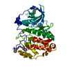
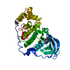
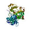
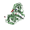
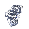
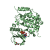
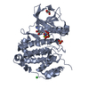
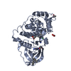
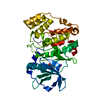
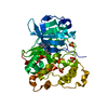
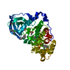
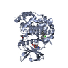
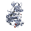
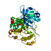
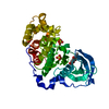
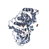
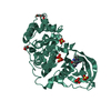
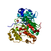
- PDB-5mo8: Crystal Structure of CK2alpha with N-(3-(((2-chloro-[1,1'-bipheny... -
+ Open data

- Basic information
| Entry | Database: PDB / ID: 5mo8 | ||||||
|---|---|---|---|---|---|---|---|
| Title | Crystal Structure of CK2alpha with N-(3-(((2-chloro-[1,1'-biphenyl]-4-yl)methyl)amino)propyl)methanesulfonamide bound | ||||||
 Components Components | Casein kinase II subunit alpha | ||||||
 Keywords Keywords | TRANSFERASE / CK2alpha / CK2a / fragment based drug discovery / high concentration screening / selective ATP competitive inhibitors / surface entrophy reduction | ||||||
| Function / homology |  Function and homology information Function and homology informationPhosphorylation and nuclear translocation of BMAL1 (ARNTL) and CLOCK / positive regulation of aggrephagy / regulation of chromosome separation / WNT mediated activation of DVL / Condensation of Prometaphase Chromosomes / protein kinase CK2 complex / symbiont-mediated disruption of host cell PML body / Phosphorylation and nuclear translocation of the CRY:PER:kinase complex / Regulation of CDH1 posttranslational processing and trafficking to plasma membrane / Receptor Mediated Mitophagy ...Phosphorylation and nuclear translocation of BMAL1 (ARNTL) and CLOCK / positive regulation of aggrephagy / regulation of chromosome separation / WNT mediated activation of DVL / Condensation of Prometaphase Chromosomes / protein kinase CK2 complex / symbiont-mediated disruption of host cell PML body / Phosphorylation and nuclear translocation of the CRY:PER:kinase complex / Regulation of CDH1 posttranslational processing and trafficking to plasma membrane / Receptor Mediated Mitophagy / Synthesis of PC / Sin3-type complex / negative regulation of signal transduction by p53 class mediator / RUNX1 interacts with co-factors whose precise effect on RUNX1 targets is not known / Maturation of hRSV A proteins / negative regulation of apoptotic signaling pathway / negative regulation of double-strand break repair via homologous recombination / positive regulation of Wnt signaling pathway / negative regulation of proteasomal ubiquitin-dependent protein catabolic process / Signal transduction by L1 / Hsp90 protein binding / PML body / Wnt signaling pathway / Regulation of PTEN stability and activity / kinase activity / positive regulation of protein catabolic process / KEAP1-NFE2L2 pathway / rhythmic process / double-strand break repair / Cooperation of PDCL (PhLP1) and TRiC/CCT in G-protein beta folding / protein folding / positive regulation of cell growth / Regulation of TP53 Activity through Phosphorylation / non-specific serine/threonine protein kinase / regulation of cell cycle / negative regulation of translation / protein stabilization / protein serine kinase activity / protein serine/threonine kinase activity / apoptotic process / positive regulation of cell population proliferation / DNA damage response / signal transduction / nucleoplasm / ATP binding / identical protein binding / nucleus / plasma membrane / cytosol Similarity search - Function | ||||||
| Biological species |  Homo sapiens (human) Homo sapiens (human) | ||||||
| Method |  X-RAY DIFFRACTION / SYNCHROTRON / MOLECULAR REPLACEMENT / molecular replacement / Resolution: 1.82 Å X-RAY DIFFRACTION / SYNCHROTRON / MOLECULAR REPLACEMENT / molecular replacement / Resolution: 1.82 Å | ||||||
 Authors Authors | Brear, P. / De Fusco, C. / Georgiou, K. / Iegre, J. / Sore, H. / Hyvonen, M. / Spring, D. | ||||||
| Funding support |  United Kingdom, 1items United Kingdom, 1items
| ||||||
 Citation Citation | Journal: Bioorg. Med. Chem. / Year: 2017 Title: A fragment-based approach leading to the discovery of a novel binding site and the selective CK2 inhibitor CAM4066. Authors: De Fusco, C. / Brear, P. / Iegre, J. / Georgiou, K.H. / Sore, H.F. / Hyvonen, M. / Spring, D.R. | ||||||
| History |
|
- Structure visualization
| Structure viewer | Molecule: MolmilJmol/JSmol |
|---|
- Downloads & links
-Download
| PDBx/mmCIF format | 5mo8.cif.gz | 293.8 KB | Display | PDBx/mmCIF format |
|---|---|---|---|---|
| PDB format | pdb5mo8.ent.gz | 240.4 KB | Display | PDB format |
| PDBx/mmJSON format | 5mo8.json.gz | Tree view | PDBx/mmJSON format | |
| Others |  Other downloads Other downloads |
-Validation report
| Arichive directory | https://data.pdbj.org/pub/pdb/validation_reports/mo/5mo8ftp://data.pdbj.org/pub/pdb/validation_reports/mo/5mo8 | HTTPS FTP |
|---|
-Related structure data
| Related structure data | 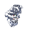 5ct0C  5ctpC  5cu0C  5cu2C 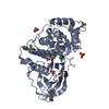 5cx9C  5mmfC  5mmrC  5mo5C  5mo6C  5mo7C  5modC  5moeC  5mohC 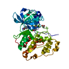 5motC  5movC  5mowC  5mp8C  5mpjC 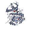 5cvhS S: Starting model for refinement C: citing same article ( |
|---|---|
| Similar structure data |


-Links
 PDBj
PDBj


- Assembly
| Deposited unit | 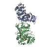
| ||||||||
|---|---|---|---|---|---|---|---|---|---|
| 1 | 
| ||||||||
| 2 | 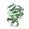
| ||||||||
| Unit cell |
|
-Components
| #1: Protein | Mass: 41467.793 Da / Num. of mol.: 2 Fragment: residues 2-329 and N-terminal extension GSMDIEFDDDADDDGSGSGSGSGS Mutation: R21S Source method: isolated from a genetically manipulated source Source: (gene. exp.) Homo sapiens (human) / Gene: CSNK2A1, CK2A1 / Production host:  References: UniProt: P68400, non-specific serine/threonine protein kinase #2: Chemical |   Mass: 59.044 Da / Num. of mol.: 3 / Source method: obtained synthetically / Formula: C2H3O2 Mass: 59.044 Da / Num. of mol.: 3 / Source method: obtained synthetically / Formula: C2H3O2#3: Chemical | 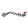  Mass: 479.955 Da / Num. of mol.: 2 / Source method: obtained synthetically / Formula: C26H26ClN3O4 Mass: 479.955 Da / Num. of mol.: 2 / Source method: obtained synthetically / Formula: C26H26ClN3O4#4: Chemical |   Mass: 94.971 Da / Num. of mol.: 2 / Source method: obtained synthetically / Formula: PO4 Mass: 94.971 Da / Num. of mol.: 2 / Source method: obtained synthetically / Formula: PO4#5: Water | ChemComp-HOH / |  Mass: 18.015 Da / Num. of mol.: 211 / Source method: isolated from a natural source / Formula: H2O Mass: 18.015 Da / Num. of mol.: 211 / Source method: isolated from a natural source / Formula: H2O |
|---|
-Experimental details
-Experiment
| Experiment | Method: X-RAY DIFFRACTION / Number of used crystals: 1 |
|---|
- Sample preparation
| Crystal | Density Matthews: 2.2 Å3/Da / Density % sol: 44.11 % / Mosaicity: 0.12 ° |
|---|---|
| Crystal grow | Temperature: 298 K / Method: vapor diffusion, hanging drop / pH: 6.5 Details: 112.5mM Mes pH 6.5, 35% glycerol ethoxylate, 180 mM ammonium acetate |
-Data collection
| Diffraction | Mean temperature: 100 K |
|---|---|
| Diffraction source | Source: SYNCHROTRON / Site: Diamond / Beamline: I03 / Wavelength: 0.97625 Å |
| Detector | Type: DECTRIS PILATUS3 6M / Detector: PIXEL / Date: Sep 17, 2015 |
| Radiation | Protocol: SINGLE WAVELENGTH / Monochromatic (M) / Laue (L): M / Scattering type: x-ray |
| Radiation wavelength | Wavelength: 0.97625 Å / Relative weight: 1 |
| Reflection | Resolution: 1.82→167.46 Å / Num. obs: 66762 / % possible obs: 100 % / Redundancy: 6.6 % / Biso Wilson estimate: 32.89 Å2 / CC1/2: 0.999 / Rmerge(I) obs: 0.058 / Rsym value: 0.058 / Net I/σ(I): 16.1 |
| Reflection shell | Resolution: 1.82→1.822 Å / Redundancy: 6.4 % / Rmerge(I) obs: 0.759 / Mean I/σ(I) obs: 2.3 / CC1/2: 0.948 / % possible all: 99.8 |
-Phasing
| Phasing | Method: molecular replacement |
|---|
- Processing
| Software |
| ||||||||||||||||||||||||||||||||||||||||||||||||||||||||||||||||||||||||||||||||||||||||||||||||||||||||||||
|---|---|---|---|---|---|---|---|---|---|---|---|---|---|---|---|---|---|---|---|---|---|---|---|---|---|---|---|---|---|---|---|---|---|---|---|---|---|---|---|---|---|---|---|---|---|---|---|---|---|---|---|---|---|---|---|---|---|---|---|---|---|---|---|---|---|---|---|---|---|---|---|---|---|---|---|---|---|---|---|---|---|---|---|---|---|---|---|---|---|---|---|---|---|---|---|---|---|---|---|---|---|---|---|---|---|---|---|---|---|
| Refinement | Method to determine structure: MOLECULAR REPLACEMENT Starting model: 5CVH Resolution: 1.82→167.46 Å / Cor.coef. Fo:Fc: 0.9377 / Cor.coef. Fo:Fc free: 0.9323 / SU R Cruickshank DPI: 0.136 / Cross valid method: THROUGHOUT / σ(F): 0 / SU R Blow DPI: 0.134 / SU Rfree Blow DPI: 0.115 / SU Rfree Cruickshank DPI: 0.117
| ||||||||||||||||||||||||||||||||||||||||||||||||||||||||||||||||||||||||||||||||||||||||||||||||||||||||||||
| Displacement parameters | Biso max: 171.83 Å2 / Biso mean: 53.73 Å2 / Biso min: 11.75 Å2
| ||||||||||||||||||||||||||||||||||||||||||||||||||||||||||||||||||||||||||||||||||||||||||||||||||||||||||||
| Refine analyze | Luzzati coordinate error obs: 0.278 Å | ||||||||||||||||||||||||||||||||||||||||||||||||||||||||||||||||||||||||||||||||||||||||||||||||||||||||||||
| Refinement step | Cycle: final / Resolution: 1.82→167.46 Å
| ||||||||||||||||||||||||||||||||||||||||||||||||||||||||||||||||||||||||||||||||||||||||||||||||||||||||||||
| Refine LS restraints |
| ||||||||||||||||||||||||||||||||||||||||||||||||||||||||||||||||||||||||||||||||||||||||||||||||||||||||||||
| LS refinement shell | Resolution: 1.82→1.86 Å / Rfactor Rfree error: 0
| ||||||||||||||||||||||||||||||||||||||||||||||||||||||||||||||||||||||||||||||||||||||||||||||||||||||||||||
| Refinement TLS params. | Method: refined / Refine-ID: X-RAY DIFFRACTION
| ||||||||||||||||||||||||||||||||||||||||||||||||||||||||||||||||||||||||||||||||||||||||||||||||||||||||||||
| Refinement TLS group |
|
