 Movie
Movie Controller

[English] 日本語
 Yorodumi
- PDB-5l47: X-ray structure of the 2-22' locally-closed mutant of GLIC in com... -
+ Open data

- Basic information
| Entry | Database: PDB / ID: 5l47 | ||||||
|---|---|---|---|---|---|---|---|
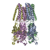
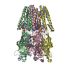
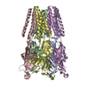
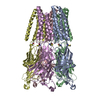
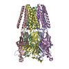
| Title | X-ray structure of the 2-22' locally-closed mutant of GLIC in complex with cyanoselenobarbital (seleniated barbiturate) | ||||||
 Components Components | Proton-gated ion channel | ||||||
 Keywords Keywords | MEMBRANE PROTEIN / TRANSPORT PROTEIN | ||||||
| Function / homology |  Function and homology information Function and homology informationsodium channel activity / potassium channel activity / extracellular ligand-gated monoatomic ion channel activity / transmembrane signaling receptor activity / identical protein binding / plasma membrane Similarity search - Function | ||||||
| Biological species |  Gloeobacter (bacteria) Gloeobacter (bacteria) | ||||||
| Method |  X-RAY DIFFRACTION / SYNCHROTRON / MOLECULAR REPLACEMENT / Resolution: 3.3 Å X-RAY DIFFRACTION / SYNCHROTRON / MOLECULAR REPLACEMENT / Resolution: 3.3 Å | ||||||
 Authors Authors | Reinholds Ruza, R. / Fourati, Z. / Delarue, M. | ||||||
| Funding support |  France, 1items France, 1items
| ||||||
 Citation Citation | Journal: J. Biol. Chem. / Year: 2017 Title: Barbiturates Bind in the GLIC Ion Channel Pore and Cause Inhibition by Stabilizing a Closed State. Authors: Fourati, Z. / Ruza, R.R. / Laverty, D. / Drege, E. / Delarue-Cochin, S. / Joseph, D. / Koehl, P. / Smart, T. / Delarue, M. | ||||||
| History |
|
- Structure visualization
| Structure viewer | Molecule: MolmilJmol/JSmol |
|---|
- Downloads & links
-Download
| PDBx/mmCIF format | 5l47.cif.gz | 318.6 KB | Display | PDBx/mmCIF format |
|---|---|---|---|---|
| PDB format | pdb5l47.ent.gz | 261.2 KB | Display | PDB format |
| PDBx/mmJSON format | 5l47.json.gz | Tree view | PDBx/mmJSON format | |
| Others |  Other downloads Other downloads |
-Validation report
| Arichive directory | https://data.pdbj.org/pub/pdb/validation_reports/l4/5l47ftp://data.pdbj.org/pub/pdb/validation_reports/l4/5l47 | HTTPS FTP |
|---|
-Related structure data
| Related structure data |  5l4eC  5l4hC  3tlvS S: Starting model for refinement C: citing same article ( |
|---|---|
| Similar structure data |
-Links
 PDBj
PDBj

- Assembly
| Deposited unit | 
| ||||||||
|---|---|---|---|---|---|---|---|---|---|
| 1 |
| ||||||||
| Unit cell |
|
-Components
-Protein , 1 types, 5 molecules ABCDE
| #1: Protein | Mass: 36225.605 Da / Num. of mol.: 5 Source method: isolated from a genetically manipulated source Source: (gene. exp.) Gloeobacter (bacteria) / Strain: PCC 7421 / Tissue: Memrane / Gene: glvI, glr4197 / Production host: |
|---|
-Non-polymers , 6 types, 123 molecules 


| #2: Chemical | ChemComp-CL /  Mass: 35.453 Da / Num. of mol.: 5 / Source method: obtained synthetically / Formula: Cl Mass: 35.453 Da / Num. of mol.: 5 / Source method: obtained synthetically / Formula: Cl#3: Chemical | ChemComp-ACT /  Mass: 59.044 Da / Num. of mol.: 5 / Source method: obtained synthetically / Formula: C2H3O2 Mass: 59.044 Da / Num. of mol.: 5 / Source method: obtained synthetically / Formula: C2H3O2#4: Chemical | ChemComp-NA /  Mass: 22.990 Da / Num. of mol.: 4 / Source method: obtained synthetically / Formula: Na Mass: 22.990 Da / Num. of mol.: 4 / Source method: obtained synthetically / Formula: Na#5: Chemical | ChemComp-6JA / |  Mass: 288.162 Da / Num. of mol.: 1 / Source method: obtained synthetically / Formula: C9H11N3O3Se Mass: 288.162 Da / Num. of mol.: 1 / Source method: obtained synthetically / Formula: C9H11N3O3Se#6: Chemical | ChemComp-D12 / |  Mass: 170.335 Da / Num. of mol.: 1 / Source method: obtained synthetically / Formula: C12H26 Mass: 170.335 Da / Num. of mol.: 1 / Source method: obtained synthetically / Formula: C12H26#7: Water | ChemComp-HOH / | Mass: 18.015 Da / Num. of mol.: 107 / Source method: isolated from a natural source / Formula: H2O |
|---|
-Details
| Has protein modification | Y |
|---|
-Experimental details
-Experiment
| Experiment | Method: X-RAY DIFFRACTION / Number of used crystals: 1 |
|---|
- Sample preparation
| Crystal | Density Matthews: 5.07 Å3/Da / Density % sol: 75.73 % |
|---|---|
| Crystal grow | Temperature: 291 K / Method: vapor diffusion, hanging drop / pH: 4 Details: 100 mM Na Acetate pH4, 200 mM Na SCN, 12-15% PEG4000, 3% DMSO, 16% glycerol PH range: 4-5 |
-Data collection
| Diffraction | Mean temperature: 80 K |
|---|---|
| Diffraction source | Source: SYNCHROTRON / Site: SOLEIL / Beamline: PROXIMA 1 / Wavelength: 0.93 Å |
| Detector | Type: DECTRIS PILATUS 6M-F / Detector: PIXEL / Date: Nov 7, 2015 |
| Radiation | Protocol: SINGLE WAVELENGTH / Monochromatic (M) / Laue (L): M / Scattering type: x-ray |
| Radiation wavelength | Wavelength: 0.93 Å / Relative weight: 1 |
| Reflection | Resolution: 2.99→50 Å / Num. obs: 72625 / % possible obs: 99.1 % / Redundancy: 3.9 % / Biso Wilson estimate: 93.49 Å2 / Net I/σ(I): 9.4 |
| Reflection shell | Resolution: 2.99→3.15 Å / Redundancy: 3.1 % / Mean I/σ(I) obs: 1.9 / % possible all: 99.5 |
- Processing
| Software |
| ||||||||||||||||||||||||||||||||||||||||||||||||||||||||||||||||||||||||||||||||||||||||||||||||||||||||||||||||||
|---|---|---|---|---|---|---|---|---|---|---|---|---|---|---|---|---|---|---|---|---|---|---|---|---|---|---|---|---|---|---|---|---|---|---|---|---|---|---|---|---|---|---|---|---|---|---|---|---|---|---|---|---|---|---|---|---|---|---|---|---|---|---|---|---|---|---|---|---|---|---|---|---|---|---|---|---|---|---|---|---|---|---|---|---|---|---|---|---|---|---|---|---|---|---|---|---|---|---|---|---|---|---|---|---|---|---|---|---|---|---|---|---|---|---|---|
| Refinement | Method to determine structure: MOLECULAR REPLACEMENT Starting model: 3TLV.pdb Resolution: 3.3→49 Å / Cor.coef. Fo:Fc: 0.8344 / Cor.coef. Fo:Fc free: 0.8028 / SU R Cruickshank DPI: 0.607 / Cross valid method: THROUGHOUT / SU R Blow DPI: 0.535 / SU Rfree Blow DPI: 0.3 / SU Rfree Cruickshank DPI: 0.313
| ||||||||||||||||||||||||||||||||||||||||||||||||||||||||||||||||||||||||||||||||||||||||||||||||||||||||||||||||||
| Displacement parameters | Biso mean: 89.93 Å2
| ||||||||||||||||||||||||||||||||||||||||||||||||||||||||||||||||||||||||||||||||||||||||||||||||||||||||||||||||||
| Refine analyze | Luzzati coordinate error obs: 0.448 Å | ||||||||||||||||||||||||||||||||||||||||||||||||||||||||||||||||||||||||||||||||||||||||||||||||||||||||||||||||||
| Refinement step | Cycle: 1 / Resolution: 3.3→49 Å
| ||||||||||||||||||||||||||||||||||||||||||||||||||||||||||||||||||||||||||||||||||||||||||||||||||||||||||||||||||
| Refine LS restraints |
| ||||||||||||||||||||||||||||||||||||||||||||||||||||||||||||||||||||||||||||||||||||||||||||||||||||||||||||||||||
| LS refinement shell | Resolution: 3.1→3.18 Å / Total num. of bins used: 20
|
