 Movie
Movie Controller

[English] 日本語
 Yorodumi
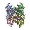
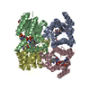
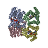
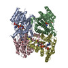
- PDB-5k6a: Trypanosoma brucei Pteridine reductase 1 (PTR1) in complex with c... -
+ Open data

- Basic information
| Entry | Database: PDB / ID: 5k6a | ||||||
|---|---|---|---|---|---|---|---|
| Title | Trypanosoma brucei Pteridine reductase 1 (PTR1) in complex with compound 1 | ||||||
 Components Components | (Pteridine reductase) x 2 | ||||||
 Keywords Keywords | OXIDOREDUCTASE / Trypanosoma brucei / pteridine reductase | ||||||
| Function / homology |  Function and homology information Function and homology information | ||||||
| Biological species |  | ||||||
| Method |  X-RAY DIFFRACTION / SYNCHROTRON / MOLECULAR REPLACEMENT / Resolution: 1.7 Å X-RAY DIFFRACTION / SYNCHROTRON / MOLECULAR REPLACEMENT / Resolution: 1.7 Å | ||||||
 Authors Authors | Landi, G. / Pozzi, C. / Di Pisa, F. / Dello lacono, L. / Mangani, S. | ||||||
 Citation Citation | Journal: Molecules / Year: 2017 Title: Chroman-4-One Derivatives Targeting Pteridine Reductase 1 and Showing Anti-Parasitic Activity. Authors: Di Pisa, F. / Landi, G. / Dello Iacono, L. / Pozzi, C. / Borsari, C. / Ferrari, S. / Santucci, M. / Santarem, N. / Cordeiro-da-Silva, A. / Moraes, C.B. / Alcantara, L.M. / Fontana, V. / ...Authors: Di Pisa, F. / Landi, G. / Dello Iacono, L. / Pozzi, C. / Borsari, C. / Ferrari, S. / Santucci, M. / Santarem, N. / Cordeiro-da-Silva, A. / Moraes, C.B. / Alcantara, L.M. / Fontana, V. / Freitas-Junior, L.H. / Gul, S. / Kuzikov, M. / Behrens, B. / Pohner, I. / Wade, R.C. / Costi, M.P. / Mangani, S. | ||||||
| History |
|
- Structure visualization
| Structure viewer | Molecule: MolmilJmol/JSmol |
|---|
- Downloads & links
-Download
| PDBx/mmCIF format | 5k6a.cif.gz | 221.2 KB | Display | PDBx/mmCIF format |
|---|---|---|---|---|
| PDB format | pdb5k6a.ent.gz | 174.5 KB | Display | PDB format |
| PDBx/mmJSON format | 5k6a.json.gz | Tree view | PDBx/mmJSON format | |
| Others |  Other downloads Other downloads |
-Validation report
| Arichive directory | https://data.pdbj.org/pub/pdb/validation_reports/k6/5k6aftp://data.pdbj.org/pub/pdb/validation_reports/k6/5k6a | HTTPS FTP |
|---|
-Related structure data
| Related structure data | 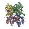 5l42C  5l4nC 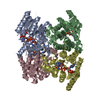 3gn1S S: Starting model for refinement C: citing same article ( |
|---|---|
| Similar structure data |


-Links
 PDBj
PDBj


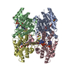
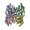
- Assembly
| Deposited unit | 
| ||||||||
|---|---|---|---|---|---|---|---|---|---|
| 1 |
| ||||||||
| Unit cell |
|
-Components
-Protein , 2 types, 4 molecules ABDC
| #1: Protein | Mass: 30685.787 Da / Num. of mol.: 3 Source method: isolated from a genetically manipulated source Source: (gene. exp.)  #2: Protein | | Mass: 30733.787 Da / Num. of mol.: 1 Source method: isolated from a genetically manipulated source Source: (gene. exp.) |
|---|
-Non-polymers , 4 types, 783 molecules 
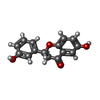


| #3: Chemical | ChemComp-NAP /  Mass: 743.405 Da / Num. of mol.: 4 / Source method: obtained synthetically / Formula: C21H28N7O17P3 Mass: 743.405 Da / Num. of mol.: 4 / Source method: obtained synthetically / Formula: C21H28N7O17P3#4: Chemical | ChemComp-6QT / (  Mass: 256.253 Da / Num. of mol.: 4 / Source method: isolated from a natural source / Formula: C15H12O4 Mass: 256.253 Da / Num. of mol.: 4 / Source method: isolated from a natural source / Formula: C15H12O4#5: Chemical |  Mass: 59.044 Da / Num. of mol.: 2 / Source method: obtained synthetically / Formula: C2H3O2 Mass: 59.044 Da / Num. of mol.: 2 / Source method: obtained synthetically / Formula: C2H3O2#6: Water | ChemComp-HOH / | Mass: 18.015 Da / Num. of mol.: 773 / Source method: isolated from a natural source / Formula: H2O |
|---|
-Experimental details
-Experiment
| Experiment | Method: X-RAY DIFFRACTION / Number of used crystals: 1 |
|---|
- Sample preparation
| Crystal | Density Matthews: 2.04 Å3/Da / Density % sol: 39.61 % |
|---|---|
| Crystal grow | Temperature: 298 K / Method: vapor diffusion Details: Protein solution: 6 g/L in 20 mM Tris-HCl pH 7.5, 10 mM DTT; Crystallization buffer: 0.1 M sodium citrate pH 5, 2.25 M sodium acetate PH range: 5.0-6.0 / Temp details: Room temperature |
-Data collection
| Diffraction | Mean temperature: 100 K |
|---|---|
| Diffraction source | Source: SYNCHROTRON / Site: ELETTRA  / Beamline: 5.2R / Wavelength: 0.999 Å / Beamline: 5.2R / Wavelength: 0.999 Å |
| Detector | Type: DECTRIS PILATUS 2M / Detector: PIXEL / Date: Aug 5, 2014 |
| Radiation | Monochromator: Si(111) / Protocol: SINGLE WAVELENGTH / Monochromatic (M) / Laue (L): M / Scattering type: x-ray |
| Radiation wavelength | Wavelength: 0.999 Å / Relative weight: 1 |
| Reflection | Resolution: 1.7→74.47 Å / Num. obs: 95660 / % possible obs: 92.66 % / Observed criterion σ(I): 2 / Redundancy: 2.3 % / Biso Wilson estimate: 8.864 Å2 / Rmerge(I) obs: 0.085 / Net I/σ(I): 6.7 |
| Reflection shell | Resolution: 1.7→1.742 Å / Redundancy: 2.2 % / Rmerge(I) obs: 0.322 / Mean I/σ(I) obs: 1.8 / % possible all: 93.5 |
- Processing
| Software |
| ||||||||||||||||||||||||||||||||||||||||||||||||||||||||||||||||||||||||||||||||||||||||||||||||||||||||||||||||||||||||||||||||||||||||||||||||||||||||||||||||||||||||||||||||||||||
|---|---|---|---|---|---|---|---|---|---|---|---|---|---|---|---|---|---|---|---|---|---|---|---|---|---|---|---|---|---|---|---|---|---|---|---|---|---|---|---|---|---|---|---|---|---|---|---|---|---|---|---|---|---|---|---|---|---|---|---|---|---|---|---|---|---|---|---|---|---|---|---|---|---|---|---|---|---|---|---|---|---|---|---|---|---|---|---|---|---|---|---|---|---|---|---|---|---|---|---|---|---|---|---|---|---|---|---|---|---|---|---|---|---|---|---|---|---|---|---|---|---|---|---|---|---|---|---|---|---|---|---|---|---|---|---|---|---|---|---|---|---|---|---|---|---|---|---|---|---|---|---|---|---|---|---|---|---|---|---|---|---|---|---|---|---|---|---|---|---|---|---|---|---|---|---|---|---|---|---|---|---|---|---|
| Refinement | Method to determine structure: MOLECULAR REPLACEMENT Starting model: 3gn1 Resolution: 1.7→74.47 Å / Cor.coef. Fo:Fc: 0.958 / Cor.coef. Fo:Fc free: 0.936 / SU B: 2.999 / SU ML: 0.093 / Cross valid method: THROUGHOUT / ESU R: 0.117 / ESU R Free: 0.115 / Stereochemistry target values: MAXIMUM LIKELIHOOD
| ||||||||||||||||||||||||||||||||||||||||||||||||||||||||||||||||||||||||||||||||||||||||||||||||||||||||||||||||||||||||||||||||||||||||||||||||||||||||||||||||||||||||||||||||||||||
| Solvent computation | Ion probe radii: 0.8 Å / Shrinkage radii: 0.8 Å / VDW probe radii: 1.2 Å / Solvent model: MASK | ||||||||||||||||||||||||||||||||||||||||||||||||||||||||||||||||||||||||||||||||||||||||||||||||||||||||||||||||||||||||||||||||||||||||||||||||||||||||||||||||||||||||||||||||||||||
| Displacement parameters | Biso mean: 18.612 Å2
| ||||||||||||||||||||||||||||||||||||||||||||||||||||||||||||||||||||||||||||||||||||||||||||||||||||||||||||||||||||||||||||||||||||||||||||||||||||||||||||||||||||||||||||||||||||||
| Refinement step | Cycle: LAST / Resolution: 1.7→74.47 Å
| ||||||||||||||||||||||||||||||||||||||||||||||||||||||||||||||||||||||||||||||||||||||||||||||||||||||||||||||||||||||||||||||||||||||||||||||||||||||||||||||||||||||||||||||||||||||
| Refine LS restraints |
|